Wang Wenxing, Wang Peiyuan, Tang Xueting, Elzatahry Ahmed A, Wang Shuwen, Al-Dahyan Daifallah, Zhao Mengyao, Yao Chi, Hung Chin-Te, Zhu Xiaohang, Zhao Tiancong, Li Xiaomin, Zhang Fan, Zhao Dongyuan
Department of Chemistry, Collaborative Innovation Center of Chemistry for Energy Materials, State Key Laboratory of Molecular Engineering of Polymers, Fudan University, Shanghai 200433, P. R. China.
Materials Science and Tech Program, College of Arts and Sciences, Qatar University, P.O. Box 2713, Doha, Qatar.
ACS Cent Sci. 2017 Aug 23;3(8):839-846. doi: 10.1021/acscentsci.7b00257. Epub 2017 Jul 26.
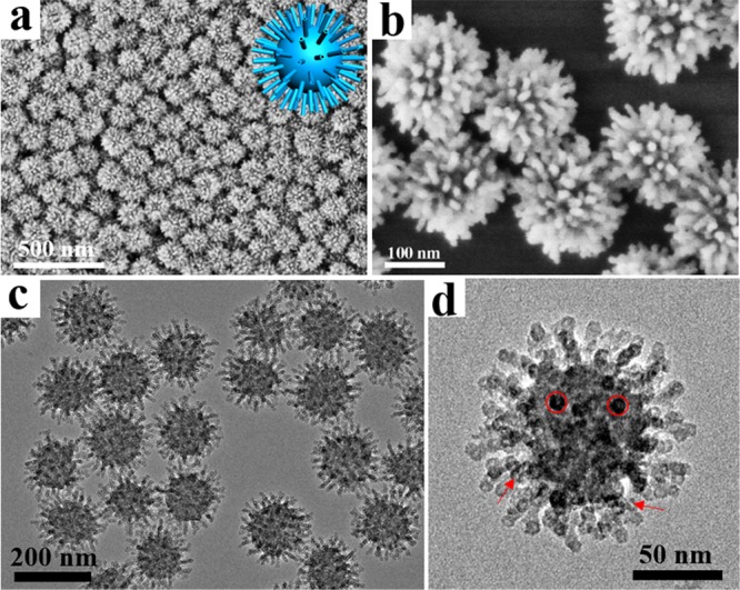
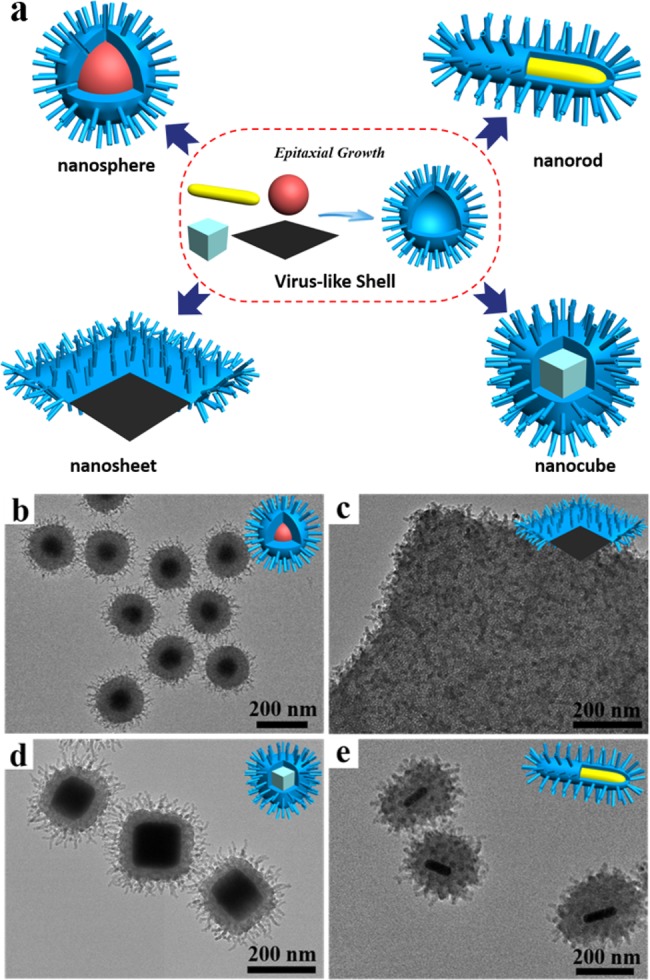
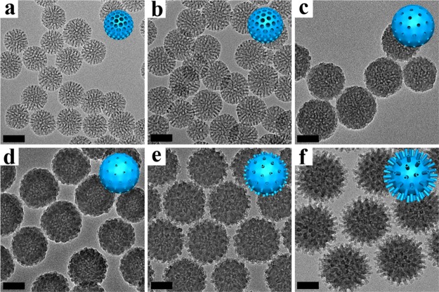
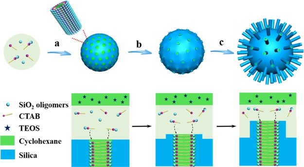
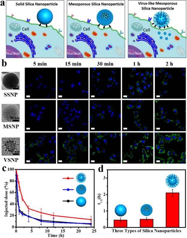
The low-efficiency cellular uptake property of current nanoparticles greatly restricts their application in the biomedical field. Herein, we demonstrate that novel virus-like mesoporous silica nanoparticles can easily be synthesized, showing greatly superior cellular uptake property. The unique virus-like mesoporous silica nanoparticles with a spiky tubular rough surface have been successfully synthesized via a novel single-micelle epitaxial growth approach in a low-concentration-surfactant oil/water biphase system. The virus-like nanoparticles' rough surface morphology results mainly from the mesoporous silica nanotubes spontaneously grown via an epitaxial growth process. The obtained nanoparticles show uniform particle size and excellent monodispersity. The structural parameters of the nanoparticles can be well tuned with controllable core diameter (∼60-160 nm), tubular length (∼6-70 nm), and outer diameter (∼6-10 nm). Thanks to the biomimetic morphology, the virus-like nanoparticles show greatly superior cellular uptake property (invading living cells in large quantities within few minutes, <5 min), unique internalization pathways, and extended blood circulation duration ( = 2.16 h), which is much longer than that of conventional mesoporous silica nanoparticles (0.45 h). Furthermore, our epitaxial growth strategy can be applied to fabricate various virus-like mesoporous core-shell structures, paving the way toward designed synthesis of virus-like nanocomposites for biomedicine applications.
当前纳米颗粒的低效率细胞摄取特性极大地限制了它们在生物医学领域的应用。在此,我们证明了新型病毒样介孔二氧化硅纳米颗粒能够轻松合成,表现出极其优异的细胞摄取特性。通过一种新颖的单胶束外延生长方法,在低浓度表面活性剂油/水双相体系中成功合成了具有尖刺状管状粗糙表面的独特病毒样介孔二氧化硅纳米颗粒。病毒样纳米颗粒的粗糙表面形态主要源于通过外延生长过程自发生长的介孔二氧化硅纳米管。所获得的纳米颗粒粒径均匀,具有出色的单分散性。纳米颗粒的结构参数可以通过可控的核心直径(约60 - 160 nm)、管状长度(约6 - 70 nm)和外径(约6 - 10 nm)进行良好调控。得益于这种仿生形态,病毒样纳米颗粒表现出极其优异的细胞摄取特性(在几分钟内,<5分钟内大量侵入活细胞)、独特的内化途径以及延长的血液循环持续时间( = 2.16小时),这比传统介孔二氧化硅纳米颗粒的血液循环持续时间(0.45小时)长得多。此外,我们的外延生长策略可用于制造各种病毒样介孔核壳结构,为设计合成用于生物医学应用的病毒样纳米复合材料铺平了道路。