Huang Fu-Chin, Hsieh Hsin-Yi, Chang Tsung C, Su Shu-Li, Tseng Shin-Ling, Lai Yu-Hsuan, Kuo Ming-Tse
Department of Ophthalmology, National Cheng Kung University Hospital, College of Medicine, National Cheng Kung University, Tainan, Taiwan.
Department of Medical Laboratory Science and Biotechnology, College of Medicine, National Cheng Kung University, Tainan, Taiwan.
Mol Vis. 2017 Aug 24;23:614-623. eCollection 2017.
Developing a DNA dot hybridization model for diagnosing parasitic keratitis.
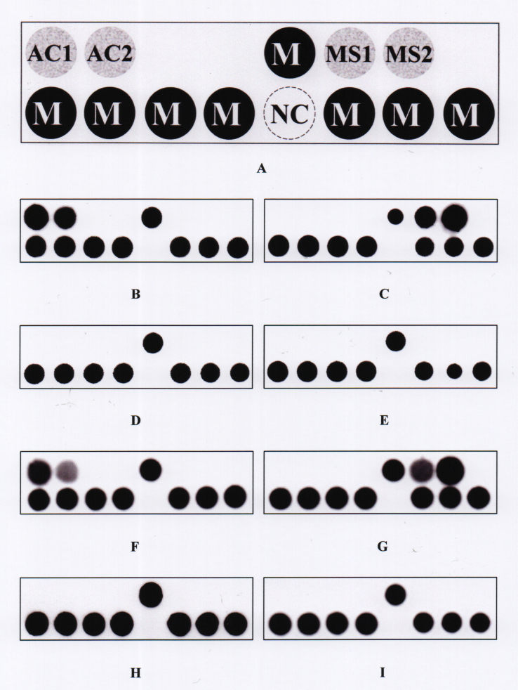
Newly designed oligonucleotide probes for detecting and microsporidia were tested with target reference strains of (n = 20) and microsporidia (n = 3), and non-target microorganisms, including bacteria (n = 20) and fungi (n = 20). These probes, which had passed the preliminary tests, were then assembled as a parasite dot hybridization (PDH) model for assessing 33 clinical samples from patients with clinically suspected and microsporidia keratitis, including eight positives for , 13 positives for microsporidia, and 12 negatives for both pathogens.
Two probes for detecting and two for detecting microsporidia passed the tests using target and non-target strains and then were assembled in the PDH model. For clinical samples, one -positive sample (proved with pathology) was falsely negative according to the PDH assay. The sensitivity and specificity of the PDH assay for diagnosing keratitis were 87.5% and 100%, respectively, while the sensitivity and specificity for diagnosing microsporidia keratitis were 100%. The infectious agent of all clinical samples of microsporidia keratitis was identified as with DNA sequencing, while those of keratitis were caused by four species of , with found in four samples (50%, 4/8).
The PDH model has the potential to be a molecular assay for diagnosing and microsporidia keratitis. However, a prospective clinical study might be needed before the model is adopted in routine clinical practice.
建立一种用于诊断寄生虫性角膜炎的DNA斑点杂交模型。
用新设计的用于检测棘阿米巴和微孢子虫的寡核苷酸探针,对棘阿米巴的目标参考菌株(n = 20)和微孢子虫的目标参考菌株(n = 3),以及包括细菌(n = 20)和真菌(n = 20)在内的非目标微生物进行检测。这些通过初步测试的探针随后被组装成寄生虫斑点杂交(PDH)模型,用于评估33例临床怀疑患有棘阿米巴和微孢子虫角膜炎患者的临床样本,其中包括8例棘阿米巴阳性、13例微孢子虫阳性以及12例两种病原体均为阴性的样本。
用于检测棘阿米巴的两种探针和用于检测微孢子虫的两种探针,在使用目标菌株和非目标菌株测试时均通过了检测,随后被组装到PDH模型中。对于临床样本,根据PDH检测,有1例棘阿米巴阳性样本(经病理学证实)为假阴性。PDH检测诊断棘阿米巴角膜炎的敏感性和特异性分别为87.5%和100%,而诊断微孢子虫角膜炎的敏感性和特异性均为100%。通过DNA测序确定所有微孢子虫角膜炎临床样本的感染病原体为棘阿米巴,而棘阿米巴角膜炎的病原体由4种棘阿米巴引起,4份样本(50%,4/8)中检测到[具体棘阿米巴种类未提及]。
PDH模型有潜力成为诊断棘阿米巴和微孢子虫角膜炎的分子检测方法。然而,在该模型被应用于常规临床实践之前,可能需要进行前瞻性临床研究。