Liu Ying, Hirayama Masatoshi, Kawakita Tetsuya, Tsubota Kazuo
Department of Ophthalmology, Keio University School of Medicine, Shinjuku, Japan.
Regulatory Biology Laboratory, Salk Institute for Biological Studies, La Jolla, CA, USA.
Stem Cells Int. 2017;2017:4923426. doi: 10.1155/2017/4923426. Epub 2017 Aug 10.
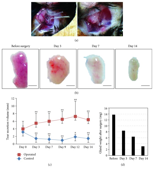
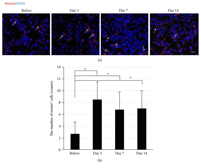
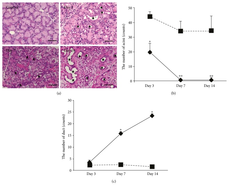
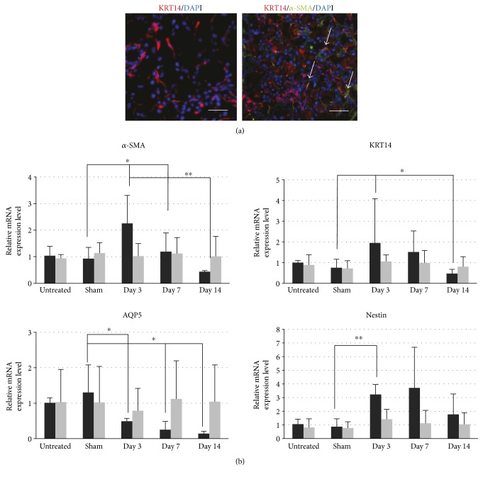
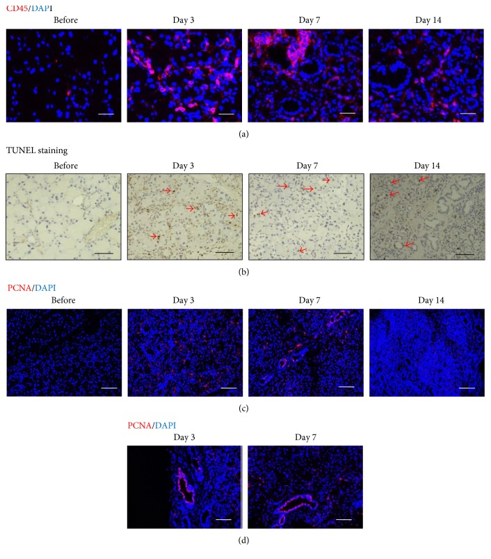
The lacrimal gland secretes tear fluids to ocular surface, which plays an indispensable role in maintaining the health of the ocular epithelia and protecting the ocular surface from the external environment. The dysfunction of the lacrimal glands causes dry eye disease due to a reduction in tear volume. The dry eye disease is becoming a popular public disease, for the number of patients is increasing, who have subjective symptom and loss of vision, which affect the quality of life. Inflammatory change in the damaged lacrimal gland has been reported; however, a major challenge is to establish a simple animal model to observe the changes. Here, we demonstrated an injury model by ligating the main excretory duct of the lacrimal gland, which is a simple and stable way to clearly understand the mechanism of lacrimal gland inflammation. We observed the process of injury and proliferation of the lacrimal gland and detected a population of lacrimal gland epithelial cells with proliferation potential which were also nestin-positive cells following duct ligation. This study successfully established an injury model to observe the tissue injury process of the lacrimal gland, and this model will be useful for analysis of the inflammation and proliferation mechanism in the future.
泪腺向眼表分泌泪液,这在维持眼表上皮健康以及保护眼表免受外部环境影响方面发挥着不可或缺的作用。泪腺功能障碍会因泪液量减少而导致干眼症。干眼症正成为一种常见的公众疾病,因为患者数量在增加,他们有主观症状和视力丧失,这会影响生活质量。已有报道泪腺受损时会发生炎症变化;然而,一个主要挑战是建立一个简单的动物模型来观察这些变化。在此,我们通过结扎泪腺的主要排泄导管展示了一种损伤模型,这是一种简单且稳定的方法,能清晰地了解泪腺炎症的机制。我们观察了泪腺损伤和增殖的过程,并检测到一群具有增殖潜能的泪腺上皮细胞,在导管结扎后这些细胞也是巢蛋白阳性细胞。本研究成功建立了一个损伤模型来观察泪腺的组织损伤过程,该模型将有助于未来对炎症和增殖机制的分析。