Department of Ophthalmology, The First Affiliated Hospital of Xiamen University, School of Medicine, Xiamen University, Xiamen, Fujian, China.
Eye Institute of Xiamen University, School of Medicine, Xiamen University, Xiamen, Fujian, China.
Invest Ophthalmol Vis Sci. 2022 Mar 2;63(3):14. doi: 10.1167/iovs.63.3.14.
To investigate microenvironment changes of the lacrimal gland after obstruction of lacrimal gland ducts.
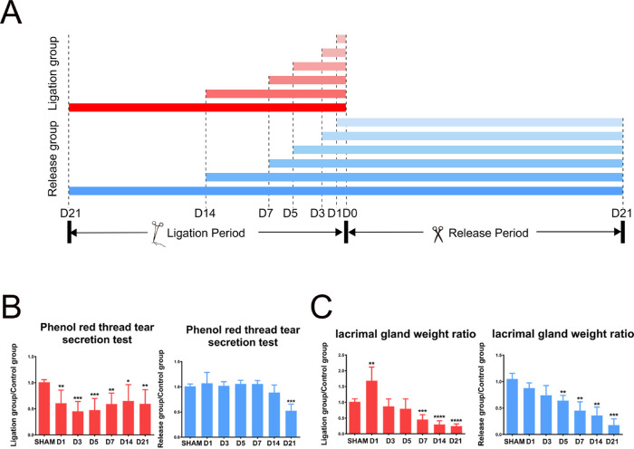
The ducts of rat exorbital lacrimal gland were ligated by sutures for different durations. After that, the sutures in some animals were released, and they were observed for 21 days to evaluate the recovery of the lacrimal gland. Slit lamp and tear secretion test was performed to evaluate ocular surface and lacrimal gland function. The lacrimal gland and cornea were harvested and processed for hematoxylin and eosin staining, oil red O staining, LipidTOX staining, Masson staining, quantitative real time polymerase chain reaction, and immunofluorescence staining.
After the lacrimal gland ducts were blocked, tear secretion and the weight of the lacrimal gland were reduced. Incidence of corneal neovascularization increased after seven days. Intraglandular ducts dilated and acini destroyed. Long-term ligation induced fibrosis and lipid accumulation of the lacrimal glands. Inflammatory cell infiltrated and inflammatory factors upregulated. Proliferative and apoptotic cells increased. Structure of myoepithelial cells and basement membrane was destroyed. The p63 expression increased whereas Pax6 expression decreased. After suture release, tear secretion and structure of acini could recover in less than seven days after ligation, with a decrease in inflammatory cell infiltration and fibrosis relief. Apoptotic cells and proliferative cells increased at five days thereafter. The structure of the myoepithelial cells and basement membrane could not recover three days after ligation, and the number of mesenchymal cells increased in ligation after five to 14 days.
Blockage of the lacrimal gland ducts results in dystrophy of lacrimal gland acini cells, inflammation, and lipid accumulation of the lacrimal gland microenvironment. Long-term duct blockage will cause irreversible lacrimal gland failure.
研究泪腺管阻塞后泪腺微环境的变化。
通过缝线结扎大鼠眶外泪腺导管不同时间,然后释放缝线,观察 21 天评估泪腺恢复情况。通过裂隙灯和泪液分泌试验评估眼表面和泪腺功能。采集泪腺和角膜进行苏木精和伊红染色、油红 O 染色、脂质染色、Masson 染色、实时定量聚合酶链反应和免疫荧光染色。
泪腺管阻塞后,泪液分泌和泪腺重量减少,第 7 天角膜新生血管增多。腺内导管扩张,腺泡破坏。长期结扎引起泪腺纤维化和脂质堆积,炎症细胞浸润,炎症因子上调,增殖和凋亡细胞增加,肌上皮细胞和基底膜结构破坏,p63 表达增加,Pax6 表达减少。释放缝线后,结扎 7 天内泪液分泌和腺泡结构可恢复,炎症细胞浸润和纤维化减轻,随后 5 天凋亡和增殖细胞增加。结扎后 3 天肌上皮细胞和基底膜结构不能恢复,5-14 天结扎后间质细胞数量增加。
泪腺管阻塞导致泪腺腺泡细胞萎缩、炎症和脂质堆积,泪腺微环境改变。长期管阻塞会导致不可逆的泪腺衰竭。