• 文献检索
  • 文档翻译
  • 深度研究
  • 学术资讯
  • Suppr Zotero 插件Zotero 插件
  • 邀请有礼
  • 套餐&价格
  • 历史记录
应用&插件
Suppr Zotero 插件Zotero 插件浏览器插件Mac 客户端Windows 客户端微信小程序
定价
高级版会员购买积分包购买API积分包
服务
文献检索文档翻译深度研究API 文档MCP 服务
关于我们
关于 Suppr公司介绍联系我们用户协议隐私条款
关注我们

Suppr 超能文献

核心技术专利:CN118964589B侵权必究
粤ICP备2023148730 号-1Suppr @ 2026

文献检索

告别复杂PubMed语法,用中文像聊天一样搜索,搜遍4000万医学文献。AI智能推荐,让科研检索更轻松。

立即免费搜索

文件翻译

保留排版,准确专业,支持PDF/Word/PPT等文件格式,支持 12+语言互译。

免费翻译文档

深度研究

AI帮你快速写综述,25分钟生成高质量综述,智能提取关键信息,辅助科研写作。

立即免费体验

啮齿动物模型中视网膜缺血/再灌注后的视神经变性

Optic Nerve Degeneration after Retinal Ischemia/Reperfusion in a Rodent Model.

作者信息

Renner Marina, Stute Gesa, Alzureiqi Mohammad, Reinhard Jacqueline, Wiemann Susanne, Schmid Heiko, Faissner Andreas, Dick H Burkhard, Joachim Stephanie C

机构信息

Experimental Eye Research, University Eye Hospital, Ruhr-University BochumBochum, Germany.

Department of Cell Morphology and Molecular Neurobiology, Faculty of Biology and Biotechnology, Ruhr-University BochumBochum, Germany.

出版信息

Front Cell Neurosci. 2017 Aug 22;11:254. doi: 10.3389/fncel.2017.00254. eCollection 2017.

DOI:10.3389/fncel.2017.00254
PMID:28878627
原文链接:https://pmc.ncbi.nlm.nih.gov/articles/PMC5572359/
Abstract

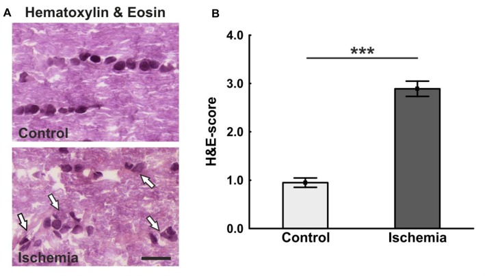
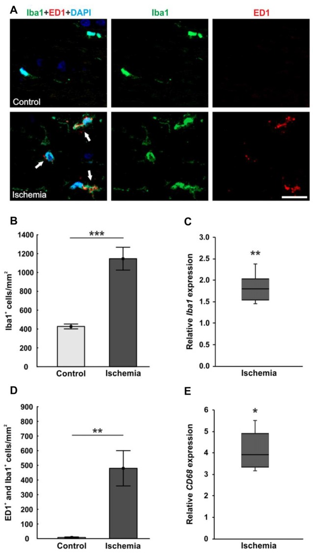
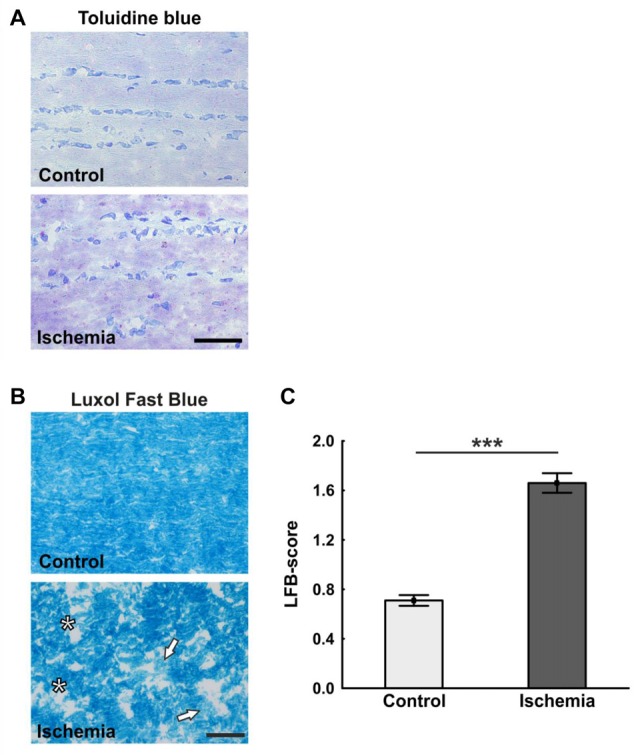
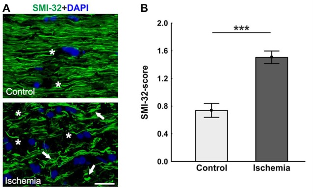
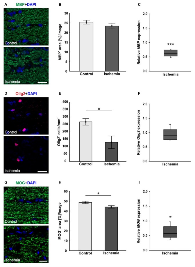
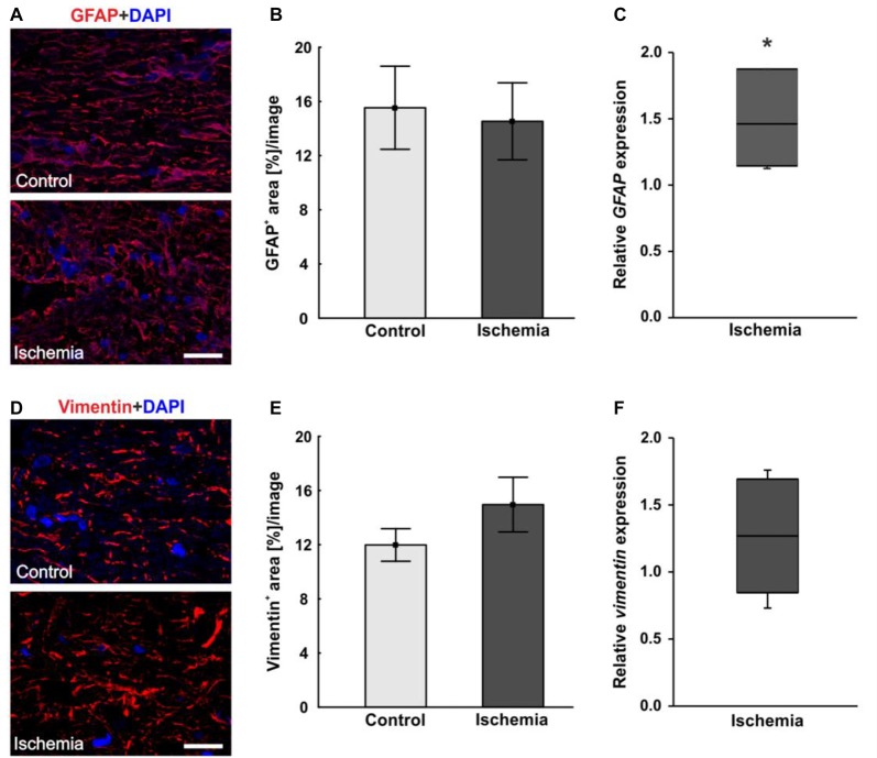
Retinal ischemia is a common pathomechanism in many ocular disorders such as age-related macular degeneration (AMD), diabetic retinopathy, glaucoma or retinal vascular occlusion. Several studies demonstrated that ischemia/reperfusion (I/R) leads to morphological and functional changes of different retinal cell types. However, little is known about the ischemic effects on the optic nerve. The goal of this study was to evaluate these effects. Ischemia was induced by raising the intraocular pressure (IOP) in one eye of rats to 140 mmHg for 1 h followed by natural reperfusion. After 21 days, histological as well as quantitative real-time PCR (qRT-PCR) analyses of optic nerves were performed. Ischemic optic nerves showed an infiltration of cells and also degeneration with signs of demyelination. Furthermore, a migration and an activation of microglia could be observed histologically as well as on mRNA level. In regard to macroglia, a trend toward gliosis could be noted after ischemia induction by vimentin staining. Additionally, an up-regulation of mRNA was found in ischemic optic nerves. Counting of oligodendrocyte transcription factor 2 positive (Olig2) cells revealed a decrease of oligodendrocytes in the ischemic group. Also, and mRNA expression was down-regulated after induction of I/R. On immunohistological level, a decrease of MOG was detectable in ischemic optic nerves as well. In addition, SMI-32 stained neurofilaments of longitudinal optic nerve sections showed a strong structural damage of the ischemic optic nerves in comparison to controls. Consequently, retinal ischemia impacts optic nerve degeneration. These findings could help to better understand the course of destruction in the optic nerve after an ischemic insult. Especially for therapeutic studies, the optic nerve is important because of its susceptibility to be damaged as a result to retinal ischemic injury and also its connecting function between the eye and the brain. So, future drug screenings should target not only the retina, but also the functionality and structure of the optic nerve. In the future, these results could lead to the development of new therapeutic strategies for treatment of ischemic injury.

摘要

视网膜缺血是许多眼部疾病如年龄相关性黄斑变性(AMD)、糖尿病性视网膜病变、青光眼或视网膜血管阻塞的常见病理机制。多项研究表明,缺血/再灌注(I/R)会导致不同视网膜细胞类型发生形态和功能变化。然而,关于缺血对视神经的影响却知之甚少。本研究的目的是评估这些影响。通过将大鼠一只眼的眼压升高至140 mmHg持续1小时,随后自然再灌注来诱导缺血。21天后,对视神经进行组织学以及定量实时PCR(qRT-PCR)分析。缺血的视神经显示出细胞浸润以及伴有脱髓鞘迹象的变性。此外,从组织学以及mRNA水平上都可观察到小胶质细胞的迁移和激活。关于大胶质细胞,通过波形蛋白染色发现在缺血诱导后有胶质增生的趋势。此外,在缺血的视神经中发现mRNA上调。少突胶质细胞转录因子2阳性(Olig2)细胞计数显示缺血组少突胶质细胞减少。同样,在I/R诱导后, 和 mRNA表达下调。在免疫组织学水平上,缺血的视神经中也可检测到髓鞘少突胶质糖蛋白(MOG)减少。此外,与对照组相比,纵向视神经切片的SMI-32染色神经丝显示缺血的视神经有严重的结构损伤。因此,视网膜缺血会影响视神经变性。这些发现有助于更好地理解缺血损伤后视神经的破坏过程。特别是对于治疗研究而言,视神经很重要,因为它容易因视网膜缺血损伤而受损,并且在眼与脑之间具有连接功能。所以,未来的药物筛选不仅应针对视网膜,还应针对视神经的功能和结构。未来,这些结果可能会导致开发出治疗缺血性损伤的新治疗策略。

https://cdn.ncbi.nlm.nih.gov/pmc/blobs/ca4e/5572359/8719fea3114a/fncel-11-00254-g0006.jpg
https://cdn.ncbi.nlm.nih.gov/pmc/blobs/ca4e/5572359/ec333613ed5f/fncel-11-00254-g0001.jpg
https://cdn.ncbi.nlm.nih.gov/pmc/blobs/ca4e/5572359/cb307e0897b6/fncel-11-00254-g0002.jpg
https://cdn.ncbi.nlm.nih.gov/pmc/blobs/ca4e/5572359/629301493195/fncel-11-00254-g0003.jpg
https://cdn.ncbi.nlm.nih.gov/pmc/blobs/ca4e/5572359/76b054ce6d64/fncel-11-00254-g0004.jpg
https://cdn.ncbi.nlm.nih.gov/pmc/blobs/ca4e/5572359/c10ba7d79f26/fncel-11-00254-g0005.jpg
https://cdn.ncbi.nlm.nih.gov/pmc/blobs/ca4e/5572359/8719fea3114a/fncel-11-00254-g0006.jpg

相似文献

1
Optic Nerve Degeneration after Retinal Ischemia/Reperfusion in a Rodent Model.啮齿动物模型中视网膜缺血/再灌注后的视神经变性
Front Cell Neurosci. 2017 Aug 22;11:254. doi: 10.3389/fncel.2017.00254. eCollection 2017.
2
Retinal ischemia triggers early microglia activation in the optic nerve followed by neurofilament degeneration.视网膜缺血引发视神经中早期小胶质细胞激活,随后出现神经丝变性。
Exp Eye Res. 2020 Sep;198:108133. doi: 10.1016/j.exer.2020.108133. Epub 2020 Jul 6.
3
Glial cell response and iNOS expression in the optic nerve head and retina of the rat following acute high IOP ischemia-reperfusion.急性高眼压缺血再灌注后大鼠视神经头和视网膜神经胶质细胞反应及诱导型一氧化氮合酶表达。
Brain Res. 2011 Jul 27;1403:67-77. doi: 10.1016/j.brainres.2011.06.005. Epub 2011 Jun 12.
4
Laquinimod protects the optic nerve and retina in an experimental autoimmune encephalomyelitis model.拉喹莫德可保护实验性自身免疫性脑脊髓炎模型中的视神经和视网膜。
J Neuroinflammation. 2018 Jun 14;15(1):183. doi: 10.1186/s12974-018-1208-3.
5
Chi-Ju-Di-Huang-Wan protects rats against retinal ischemia by downregulating matrix metalloproteinase-9 and inhibiting p38 mitogen-activated protein kinase.杞菊地黄丸通过下调基质金属蛋白酶-9和抑制p38丝裂原活化蛋白激酶来保护大鼠免受视网膜缺血损伤。
Chin Med. 2016 Sep 9;11(1):39. doi: 10.1186/s13020-016-0109-6. eCollection 2016.
6
Vascular endothelial growth factor is present in glial cells of the retina and optic nerve of human subjects with nonproliferative diabetic retinopathy.血管内皮生长因子存在于患有非增殖性糖尿病视网膜病变的人类受试者的视网膜和视神经的神经胶质细胞中。
Invest Ophthalmol Vis Sci. 1997 Jan;38(1):36-47.
7
High intraocular pressure-induced ischemia and reperfusion injury in the optic nerve and retina in rats.大鼠高眼压诱导的视神经和视网膜缺血再灌注损伤。
Graefes Arch Clin Exp Ophthalmol. 1996 Jul;234(7):445-51. doi: 10.1007/BF02539411.
8
Concentration-Dependent Inner Retina Layer Damage and Optic Nerve Degeneration in a NMDA Model.浓度依赖性内视网膜层损伤和 NMDA 模型中的视神经变性。
J Mol Neurosci. 2017 Dec;63(3-4):283-299. doi: 10.1007/s12031-017-0978-x. Epub 2017 Sep 29.
9
Glial cells in transected optic nerves of immature rats. II. An immunohistochemical study.未成熟大鼠横断视神经中的神经胶质细胞。II. 免疫组织化学研究。
J Neurocytol. 1996 Jun;25(6):381-92. doi: 10.1007/BF02284809.
10
Involvement of endoplasmic reticulum stress in optic nerve degeneration following N-methyl-D-aspartate-induced retinal damage in mice.内质网应激在 N-甲基-D-天冬氨酸诱导的小鼠视网膜损伤后视神经变性中的作用。
J Neurosci Res. 2012 Oct;90(10):1960-9. doi: 10.1002/jnr.23078. Epub 2012 Jun 5.

引用本文的文献

1
Sunitinib's Effect on Bilateral Optic Nerve Damage in Rats Following the Unilateral Clamping and Unclamping of the Common Carotid Artery.舒尼替尼对大鼠单侧颈总动脉夹闭与再夹闭后双侧视神经损伤的影响
Biomedicines. 2025 Mar 3;13(3):620. doi: 10.3390/biomedicines13030620.
2
Engineered bio-functional material-based nerve guide conduits for optic nerve regeneration: a view from the cellular perspective, challenges and the future outlook.基于工程化生物功能材料的视神经再生神经引导导管:从细胞角度、挑战及未来展望
Regen Biomater. 2024 Nov 22;12:rbae133. doi: 10.1093/rb/rbae133. eCollection 2025.
3
From Nature to Treatment: The Impact of Pterostilbene on Mitigating Retinal Ischemia-Reperfusion Damage by Reducing Oxidative Stress, Inflammation, and Apoptosis.

本文引用的文献

1
Oligodendrocyte development in the embryonic tuberal hypothalamus and the influence of Ascl1.胚胎期结节下丘脑少突胶质细胞的发育及Ascl1的影响
Neural Dev. 2016 Nov 18;11(1):20. doi: 10.1186/s13064-016-0075-9.
2
Early remodelling of the extracellular matrix proteins tenascin-C and phosphacan in retina and optic nerve of an experimental autoimmune glaucoma model.实验性自身免疫性青光眼模型视网膜和视神经中细胞外基质蛋白腱生蛋白-C和磷蛋白聚糖的早期重塑
J Cell Mol Med. 2016 Nov;20(11):2122-2137. doi: 10.1111/jcmm.12909. Epub 2016 Jul 4.
3
Simultaneous Complement Response via Lectin Pathway in Retina and Optic Nerve in an Experimental Autoimmune Glaucoma Model.
从天然产物到治疗手段:紫檀芪通过减轻氧化应激、炎症和细胞凋亡对缓解视网膜缺血再灌注损伤的影响
Life (Basel). 2024 Sep 11;14(9):1148. doi: 10.3390/life14091148.
4
Deletion of myeloid HDAC3 promotes efferocytosis to ameliorate retinal ischemic injury.删除髓系 HDAC3 可促进吞噬作用,从而改善视网膜缺血性损伤。
J Neuroinflammation. 2024 Jul 12;21(1):170. doi: 10.1186/s12974-024-03159-8.
5
Ucf-101 alleviates Ischaemia/Reperfusion induced retinal inflammation and injury via suppressing oxidative damage.Ucf-101 通过抑制氧化损伤缓解缺血/再灌注诱导的视网膜炎症和损伤。
J Mol Histol. 2024 Aug;55(4):455-464. doi: 10.1007/s10735-024-10213-5. Epub 2024 Jun 15.
6
Retrolaminar Demyelination of Structurally Intact Axons in Nonhuman Primate Experimental Glaucoma.结构完整轴突的神经髓鞘在非人类灵长类实验性青光眼的后退性脱髓鞘。
Invest Ophthalmol Vis Sci. 2024 Feb 1;65(2):36. doi: 10.1167/iovs.65.2.36.
7
Gliosis-dependent expression of complement factor H truncated variants attenuates retinal neurodegeneration following ischemic injury.胶质细胞依赖性补体因子 H 截断变体的表达减轻缺血性损伤后视网膜神经退行性变。
J Neuroinflammation. 2024 Feb 22;21(1):56. doi: 10.1186/s12974-024-03045-3.
8
Glaucoma Animal Models beyond Chronic IOP Increase.青光眼动物模型的研究进展:超越慢性眼压升高。
Int J Mol Sci. 2024 Jan 11;25(2):906. doi: 10.3390/ijms25020906.
9
Homer1 Protects against Retinal Ganglion Cell Pyroptosis by Inhibiting Endoplasmic Reticulum Stress-Associated TXNIP/NLRP3 Inflammasome Activation after Middle Cerebral Artery Occlusion-Induced Retinal Ischemia. Homer1 通过抑制脑缺血后视网膜缺血诱导的内质网应激相关 TXNIP/NLRP3 炎性小体激活来保护视网膜神经节细胞发生细胞焦亡。
Int J Mol Sci. 2023 Nov 27;24(23):16811. doi: 10.3390/ijms242316811.
10
HS Regulates the Phenotypic Transformation of Astrocytes Following Cerebral Ischemia/Reperfusion via Inhibiting the RhoA/ROCK Pathway.HS 通过抑制 RhoA/ROCK 通路调节脑缺血/再灌注后星形胶质细胞的表型转化。
Mol Neurobiol. 2024 Jun;61(6):3179-3197. doi: 10.1007/s12035-023-03797-8. Epub 2023 Nov 18.
实验性自身免疫性青光眼模型中视网膜和视神经通过凝集素途径的补体同时反应
Front Cell Neurosci. 2016 Jun 1;10:140. doi: 10.3389/fncel.2016.00140. eCollection 2016.
4
Retinal Macroglial Responses in Health and Disease.视网膜大胶质细胞在健康与疾病中的反应
Biomed Res Int. 2016;2016:2954721. doi: 10.1155/2016/2954721. Epub 2016 May 18.
5
Retinal and Optic Nerve Damage is Associated with Early Glial Responses in an Experimental Autoimmune Glaucoma Model.视网膜和视神经损伤与实验性自身免疫性青光眼模型中的早期胶质细胞反应相关。
J Mol Neurosci. 2016 Apr;58(4):470-82. doi: 10.1007/s12031-015-0707-2. Epub 2016 Jan 8.
6
Astrocytes: a central element in neurological diseases.星形胶质细胞:神经疾病的核心要素。
Acta Neuropathol. 2016 Mar;131(3):323-45. doi: 10.1007/s00401-015-1513-1. Epub 2015 Dec 15.
7
Oligodendrocyte Development and Myelination in Neurodevelopment: Molecular Mechanisms in Health and Disease.神经发育中的少突胶质细胞发育与髓鞘形成:健康与疾病中的分子机制
Curr Pharm Des. 2016;22(6):656-79. doi: 10.2174/1381612822666151204000636.
8
S100 alone has the same destructive effect on retinal ganglion cells as in combination with HSP 27 in an autoimmune glaucoma model.在自身免疫性青光眼模型中,单独的S100与联合HSP 27时对视网膜神经节细胞具有相同的破坏作用。
J Mol Neurosci. 2015 May;56(1):228-36. doi: 10.1007/s12031-014-0485-2. Epub 2015 Jan 11.
9
Toll-like receptor 3 activation drives the inflammatory response in oxygen-induced retinopathy in rats.Toll样受体3激活驱动大鼠氧诱导性视网膜病变中的炎症反应。
Br J Ophthalmol. 2015 Jan;99(1):125-32. doi: 10.1136/bjophthalmol-2014-305690. Epub 2014 Oct 29.
10
EDITORIAL Neuroglia as a Central Element of Neurological Diseases: An Underappreciated Target for Therapeutic Intervention.社论:神经胶质作为神经疾病的核心要素:治疗干预的一个未被充分认识的靶点。
Curr Neuropharmacol. 2014 Jul;12(4):303-7. doi: 10.2174/1570159X12999140829152550.