Alzheimer Center & Department of Neurology, Neuroscience Campus Amsterdam, VU University Medical Center, Amsterdam, The Netherlands.
Alzheimer Center & Department of Neurology, VU University Medical Center, PO Box 7057, 1007 MB, Amsterdam, The Netherlands.
Alzheimers Res Ther. 2017 Sep 12;9(1):73. doi: 10.1186/s13195-017-0299-x.
Amyloid pathology in subjects with mild cognitive impairment (MCI) is an important risk factor for progression to dementia due to Alzheimer's disease. Predicting the onset of dementia is challenging even in the presence of amyloid, as time to progression varies considerably among patients and depends on the onset of neurodegeneration. Survival analysis can account for variability in time to event, but has not often been applied to MRI measurements beyond singular predefined brain regions such as the hippocampus. Here we used a voxel-wise survival analysis to identify in an unbiased fashion brain regions where decreased gray matter volume is associated with time to dementia, and assessed the effects of amyloid on these associations.
We included 276 subjects with MCI (mean age 67 ± 8, 41% female, mean Mini-Mental State Examination 26.6 ± 2.4), baseline 3D T1-weighted structural MRI, baseline cerebrospinal fluid (CSF) biomarkers, and prospective clinical follow-up. We fitted for each voxel a proportional Cox hazards regression model to study whether decreased gray matter volume predicted progression to dementia in the total sample, and stratified for baseline amyloid status.
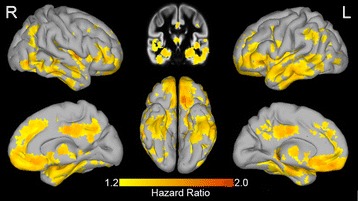
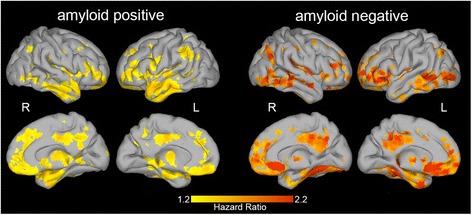
Dementia at follow-up occurred in 122 (44%) subjects over an average follow-up period of 2.5 ± 1.5 years. Baseline amyloid positivity was associated with progression to dementia (hazard ratio 2.4, p < 0.001). Within amyloid-positive subjects, decreased gray matter volume in the hippocampal, temporal, parietal, and frontal regions was associated with more rapid progression to dementia (median (interquartile range) hazard ratio across significant voxels 1.35 (1.32-1.40)). Repeating the analysis in amyloid-negative subjects revealed similar patterns (median (interquartile range) hazard ratio 1.76 (1.66-1.91)).
In subjects with MCI, both abnormal amyloid CSF and decreased gray matter volume were associated with future progression to dementia. The spatial pattern of decreased gray matter volume associated with progression to dementia was consistent for amyloid-positive and amyloid-negative subjects.
轻度认知障碍(MCI)患者的淀粉样蛋白病理学是导致阿尔茨海默病痴呆进展的重要危险因素。即使存在淀粉样蛋白,预测痴呆的发病也具有挑战性,因为患者之间的进展时间差异很大,并且取决于神经退行性变的发生。生存分析可以解释事件时间的可变性,但尚未广泛应用于 MRI 测量,超出了单一预定义的脑区,如海马体。在这里,我们使用体素生存分析以无偏倚的方式识别与痴呆发生时间相关的灰质体积减少的脑区,并评估淀粉样蛋白对这些关联的影响。
我们纳入了 276 名 MCI 患者(平均年龄 67±8 岁,41%为女性,平均简易精神状态检查 26.6±2.4 分),基线 3D T1 加权结构 MRI、基线脑脊液(CSF)生物标志物和前瞻性临床随访。我们为每个体素拟合比例 Cox 风险回归模型,以研究总样本中灰质体积减少是否预测向痴呆的进展,并按基线淀粉样蛋白状态进行分层。
在平均 2.5±1.5 年的随访期间,122 名(44%)患者出现了随访时的痴呆。基线淀粉样蛋白阳性与向痴呆的进展相关(风险比 2.4,p<0.001)。在淀粉样蛋白阳性的患者中,海马体、颞叶、顶叶和额叶区域的灰质体积减少与向痴呆的进展速度更快相关(在显著体素中,中位数(四分位距)风险比为 1.35(1.32-1.40))。在淀粉样蛋白阴性的患者中重复该分析揭示了类似的模式(中位数(四分位距)风险比为 1.76(1.66-1.91))。
在 MCI 患者中,异常的淀粉样蛋白 CSF 和灰质体积减少都与未来向痴呆的进展相关。与向痴呆进展相关的灰质体积减少的空间模式在淀粉样蛋白阳性和淀粉样蛋白阴性的患者中是一致的。