Department of Anatomy and Cell Biology, University Medicine Greifswald, Greifswald, Germany.
Department of Pathology, University Medicine Greifswald, Greifswald, Germany.
Sci Rep. 2017 Sep 13;7(1):11473. doi: 10.1038/s41598-017-11553-x.
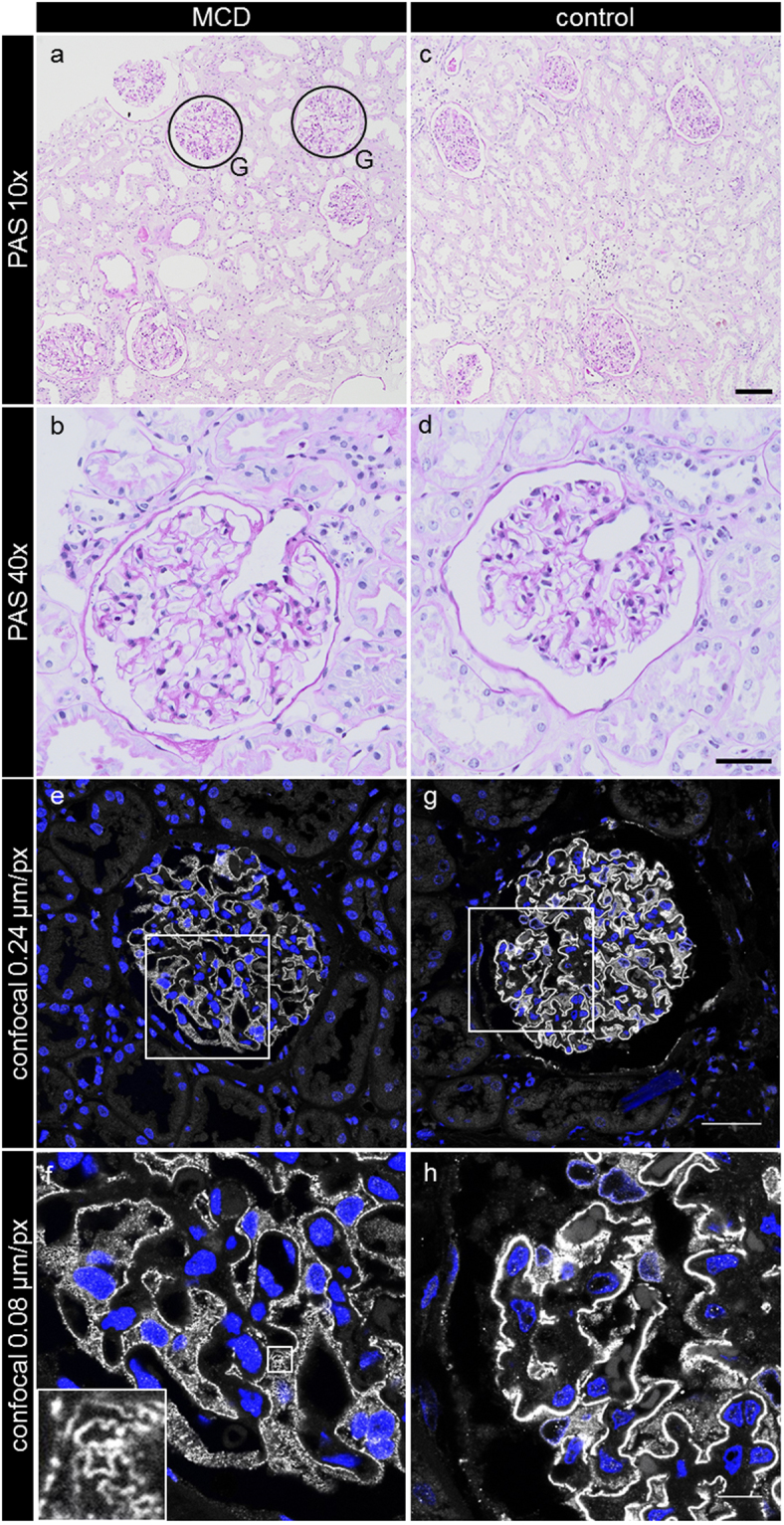
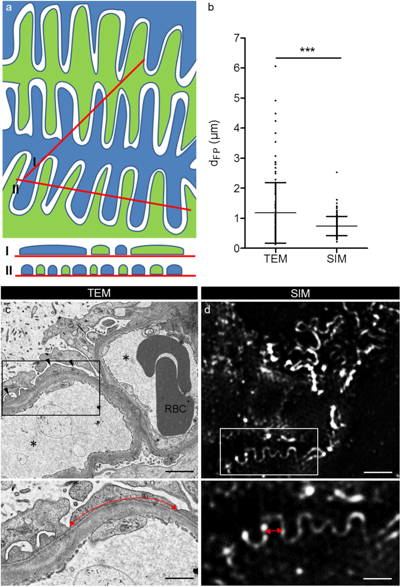
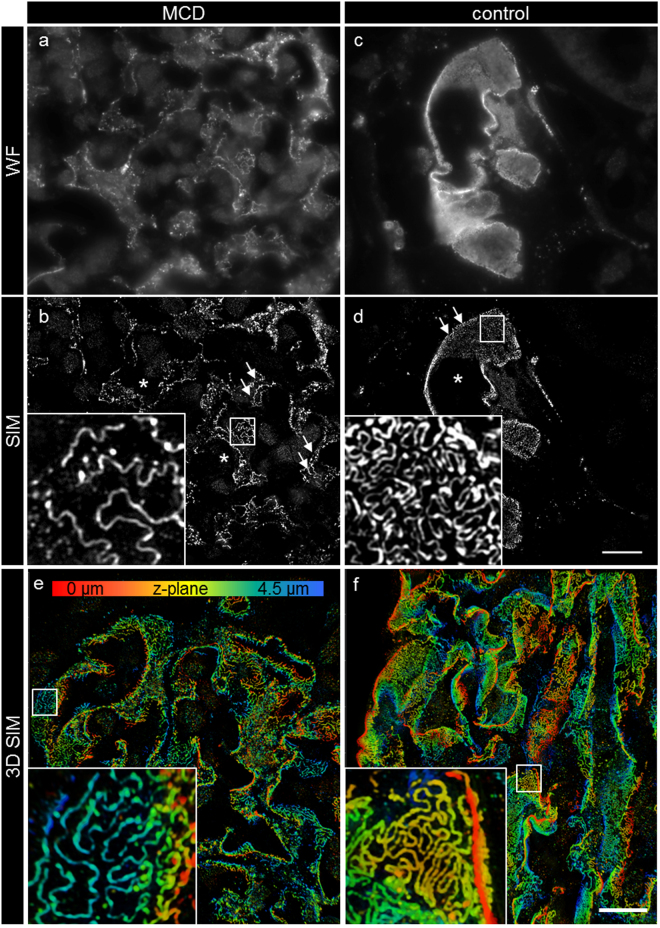
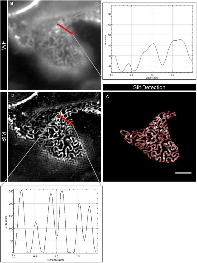
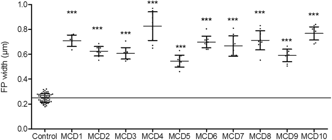
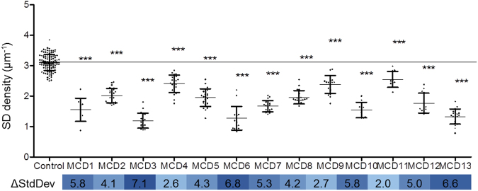
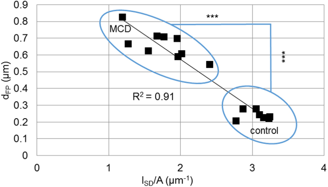
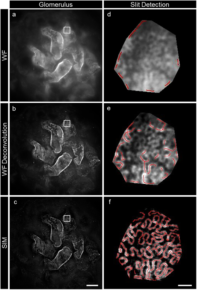
The morphology of podocyte foot processes is obligatory for renal function. Here we describe a method for the superresolution-visualization of podocyte foot processes using structured illumination microscopy of the slit diaphragm, which before has only been achieved by electron microscopy. As a proof of principle, we measured a mean foot process width of 0.249 ± 0.068 µm in healthy kidneys and a significant higher mean foot process width of 0.675 ± 0.256 µm in minimal change disease patients indicating effacement of foot processes. We then hypothesized that the slit length per glomerular capillary surface area (slit diaphragm density) could be used as an equivalent for the diagnosis of effacement. Using custom-made software we measured a mean value of 3.10 ± 0.27 µm in healthy subjects and 1.83 ± 0.49 µm in the minimal change disease patients. As foot process width was highly correlated with slit diaphragm density (R = 0.91), we concluded that our approach is a valid method for the diagnosis of foot process effacement. In summary, we present a new technique to quantify podocyte damage, which combines superresolution microscopy with automatized image processing. Due to its diverse advantages, we propose this technique to be included into routine diagnostics of glomerular histopathology.
足细胞足突的形态对于肾功能是必需的。在这里,我们描述了一种使用裂隙隔膜的结构光照明显微术对足细胞足突进行超分辨率可视化的方法,这在此之前只能通过电子显微镜来实现。作为原理的证明,我们在健康肾脏中测量到平均足突宽度为 0.249 ± 0.068 µm,而在微小病变疾病患者中测量到明显更高的平均足突宽度为 0.675 ± 0.256 µm,表明足突消失。然后我们假设,每个肾小球毛细血管表面积的裂孔长度(裂孔隔膜密度)可作为消失的诊断等效物。使用定制的软件,我们在健康受试者中测量到平均 3.10 ± 0.27 µm,在微小病变疾病患者中测量到 1.83 ± 0.49 µm。由于足突宽度与裂孔隔膜密度高度相关(R = 0.91),我们得出结论,我们的方法是诊断足突消失的有效方法。总之,我们提出了一种新的技术来定量测量足细胞损伤,该技术将超分辨率显微镜与自动化图像处理相结合。由于其多种优势,我们建议将该技术纳入肾小球组织病理学的常规诊断中。