Krieg Sandro M, Voigt Florian, Knuefermann Pascal, Kirschning Carsten Jürgen, Plesnila Nikolaus, Ringel Florian
Department of Neurosurgery, Technische Universität München, Munich, Germany.
Institute for Surgical Research, University of Munich Medical Center, Ludwig-Maximilians-Universität München, Munich, Germany.
Front Neurol. 2017 Aug 30;8:455. doi: 10.3389/fneur.2017.00455. eCollection 2017.
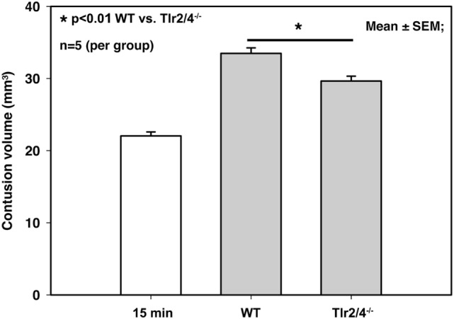
Danger-associated molecular patterns are released by damaged cells and trigger neuroinflammation through activation of non-specific pattern recognition receptors, e.g., toll-like receptors (TLRs). Since the role of TLR2 and 4 after traumatic brain injury (TBI) is still unclear, we examined the outcome and the expression of pro-inflammatory mediators after experimental TBI in and wild-type (WT) mice. and WT mice were subjected to controlled cortical injury and contusion volume and brain edema formation were assessed 24 h thereafter. Expression of inflammatory markers in brain tissue was measured by quantitative PCR 15 min, 3 h, 6 h, 12 h, and 24 h after controlled cortical impact (CCI). Contusion volume was significantly attenuated in mice (29.7 ± 0.7 mm as compared to 33.5 ± 0.8 mm in WT; < 0.05) after CCI while brain edema was not affected. Only interleukin (IL)-1β gene expression was increased after CCI in the relative to WT mice. Inducible nitric oxide synthetase, TNF, IL-6, and COX-2 were similar in injured WT and mice, while the increase in high-mobility group box 1 was attenuated at 6 h. TLR2 and 4 are consequently shown to potentially promote secondary brain injury after experimental CCI neuroinflammation and may therefore represent a novel therapeutic target for the treatment of TBI.
危险相关分子模式由受损细胞释放,并通过激活非特异性模式识别受体(如Toll样受体(TLR))触发神经炎症。由于创伤性脑损伤(TBI)后TLR2和TLR4的作用仍不清楚,我们在 小鼠和野生型(WT)小鼠中研究了实验性TBI后的结果以及促炎介质的表达。对 小鼠和WT小鼠进行控制性皮质损伤,并在24小时后评估挫伤体积和脑水肿形成情况。在控制性皮质撞击(CCI)后15分钟、3小时、6小时、12小时和24小时,通过定量PCR测量脑组织中炎症标志物的表达。CCI后, 小鼠的挫伤体积显著减小(29.7±0.7mm,而WT小鼠为33.5±0.8mm;P<0.05),而脑水肿未受影响。与WT小鼠相比,CCI后 小鼠中仅白细胞介素(IL)-1β基因表达增加。诱导型一氧化氮合酶、TNF、IL-6和COX-2在受伤的WT小鼠和 小鼠中相似,而高迁移率族蛋白B1在6小时时的增加减弱。因此,TLR2和TLR4在实验性CCI后可能通过促进神经炎症而潜在地促进继发性脑损伤,因此可能代表TBI治疗的一个新的治疗靶点。