CD Laboratory on Mechanistic and Physiological Methods for Improved Bioprocesses, TU Wien, Gumpendorferstrasse 1a/166, 1060, Vienna, Austria.
Research Area Biochemical Engineering, Institute for Chemical, Environmental and Biological Engineering, TU Wien, Gumpendorferstrasse 1a/166, 1060, Vienna, Austria.
Appl Microbiol Biotechnol. 2017 Oct;101(20):7675-7688. doi: 10.1007/s00253-017-8475-2. Epub 2017 Sep 14.
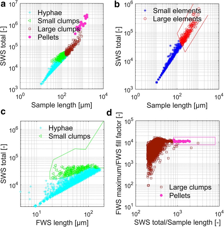
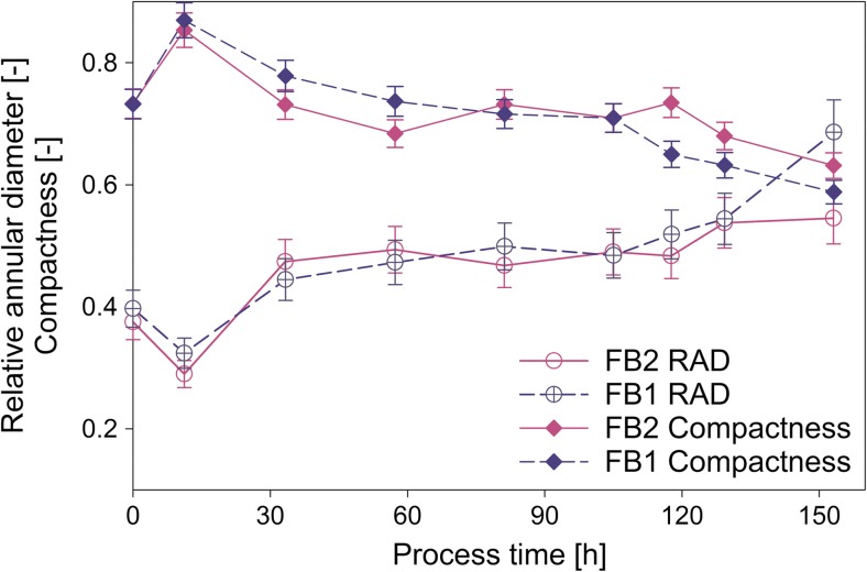
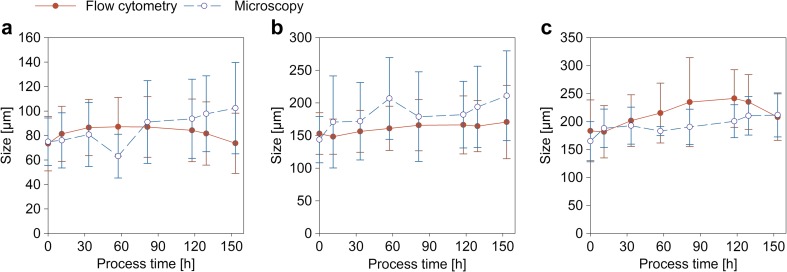
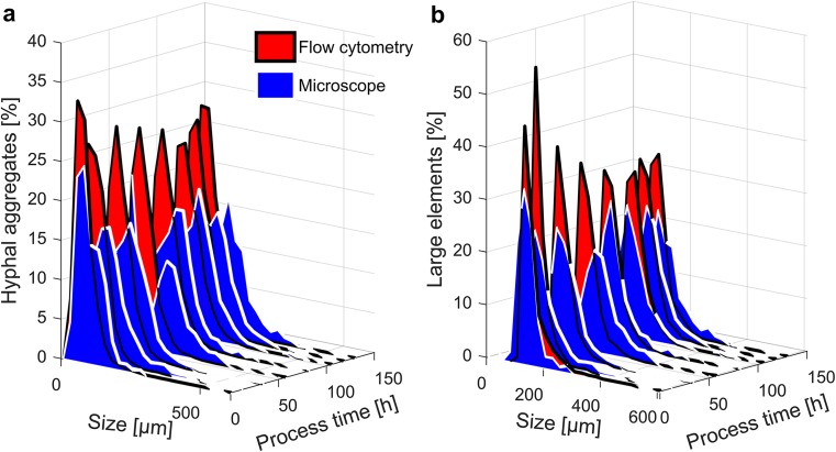
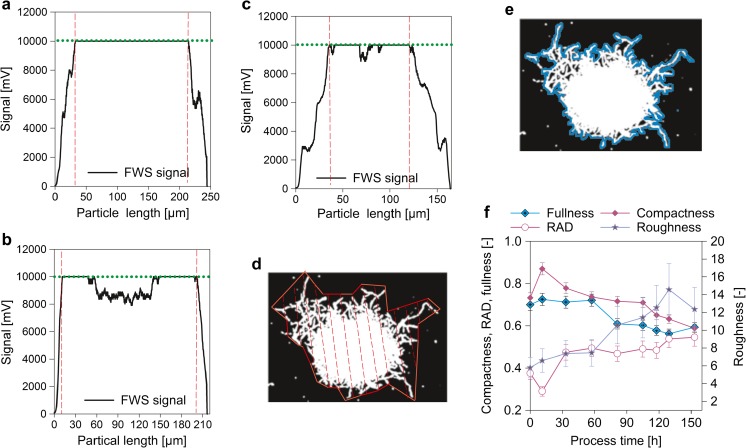
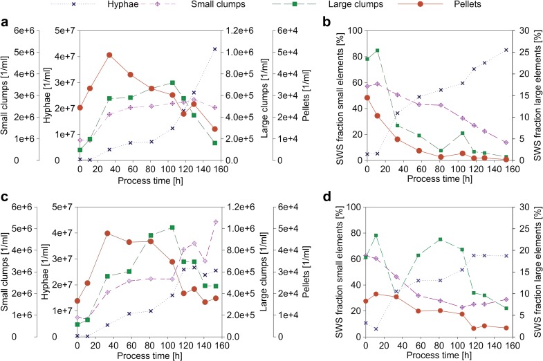
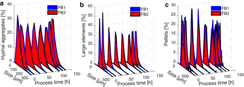
An important parameter in filamentous bioreactor cultivations is the morphology of the fungi, due to its interlink to productivity and its dependency on process conditions. Filamentous fungi show a large variety of morphological forms in submerged cultures. These range from dispersed hyphae, to interwoven mycelial aggregates, to denser hyphal aggregates, the so-called pellets. Depending on the objective function of the bioprocess, different characteristics of the morphology are favorable and need to be quantified accurately. The most common method to quantitatively characterize morphology is image analysis based on microscopy. This method is work intensive and time consuming. Therefore, we developed a faster, at-line applicable, alternative method based on flow cytometry. Within this contribution, this novel method is compared to microscopy for a penicillin production process. Both methods yielded in comparable distinction of morphological sub-populations and described their morphology in more detail. In addition to the appropriate quantification of size parameters and the description of the hyphal region around pellets, the flow cytometry method even revealed a novel compactness parameter for fungal pellets which is not accessible via light microscopy. Hence, the here presented flow cytometry method for morphological analysis is a fast and reliable alternative to common tools with some new insights in the pellet morphology, enabling at-line use in production environments.
在丝状生物反应器培养中,一个重要的参数是真菌的形态,因为它与生产力有关,并且依赖于工艺条件。丝状真菌在液体培养中表现出多种形态。这些形态从分散的菌丝体,到交织的菌丝体聚集物,到更密集的菌丝体聚集物,即所谓的颗粒。根据生物工艺的目标函数,形态的不同特征是有利的,需要准确地量化。定量描述形态最常用的方法是基于显微镜的图像分析。这种方法劳动强度大,耗时耗力。因此,我们开发了一种基于流式细胞术的更快、在线适用的替代方法。在本研究中,将这种新方法与显微镜用于青霉素生产过程进行了比较。这两种方法都能够对形态亚群进行可比的区分,并更详细地描述它们的形态。除了对大小参数进行适当的定量和描述颗粒周围菌丝区域外,流式细胞术方法甚至揭示了一种用于真菌颗粒的新的紧凑性参数,这是通过光显微镜无法获得的。因此,这里提出的用于形态分析的流式细胞术方法是一种快速可靠的替代常用工具的方法,可以提供颗粒形态的新见解,使其能够在生产环境中在线使用。