• 文献检索
  • 文档翻译
  • 深度研究
  • 学术资讯
  • Suppr Zotero 插件Zotero 插件
  • 邀请有礼
  • 套餐&价格
  • 历史记录
应用&插件
Suppr Zotero 插件Zotero 插件浏览器插件Mac 客户端Windows 客户端微信小程序
定价
高级版会员购买积分包购买API积分包
服务
文献检索文档翻译深度研究API 文档MCP 服务
关于我们
关于 Suppr公司介绍联系我们用户协议隐私条款
关注我们

Suppr 超能文献

核心技术专利:CN118964589B侵权必究
粤ICP备2023148730 号-1Suppr @ 2026

文献检索

告别复杂PubMed语法,用中文像聊天一样搜索,搜遍4000万医学文献。AI智能推荐,让科研检索更轻松。

立即免费搜索

文件翻译

保留排版,准确专业,支持PDF/Word/PPT等文件格式,支持 12+语言互译。

免费翻译文档

深度研究

AI帮你快速写综述,25分钟生成高质量综述,智能提取关键信息,辅助科研写作。

立即免费体验

智能手机上全切片成像系统的开发及冷冻切片样本评估

Development of a Whole Slide Imaging System on Smartphones and Evaluation With Frozen Section Samples.

作者信息

Yu Hong, Gao Feng, Jiang Liren, Ma Shuoxin

机构信息

Department of Pathology Center, Shanghai General Hospital, Shanghai Jiao Tong University School of Medicine (originally named "Shanghai First People's Hospital"), Shanghai, China.

TerryDr Info Technology Co., Ltd, Nanjing, Jiangsu, China.

出版信息

JMIR Mhealth Uhealth. 2017 Sep 15;5(9):e132. doi: 10.2196/mhealth.8242.

DOI:10.2196/mhealth.8242
PMID:28916508
原文链接:https://pmc.ncbi.nlm.nih.gov/articles/PMC5622289/
Abstract

BACKGROUND

The aim was to develop scalable Whole Slide Imaging (sWSI), a WSI system based on mainstream smartphones coupled with regular optical microscopes. This ultra-low-cost solution should offer diagnostic-ready imaging quality on par with standalone scanners, supporting both oil and dry objective lenses of different magnifications, and reasonably high throughput. These performance metrics should be evaluated by expert pathologists and match those of high-end scanners.

OBJECTIVE

The aim was to develop scalable Whole Slide Imaging (sWSI), a whole slide imaging system based on smartphones coupled with optical microscopes. This ultra-low-cost solution should offer diagnostic-ready imaging quality on par with standalone scanners, supporting both oil and dry object lens of different magnification. All performance metrics should be evaluated by expert pathologists and match those of high-end scanners.

METHODS

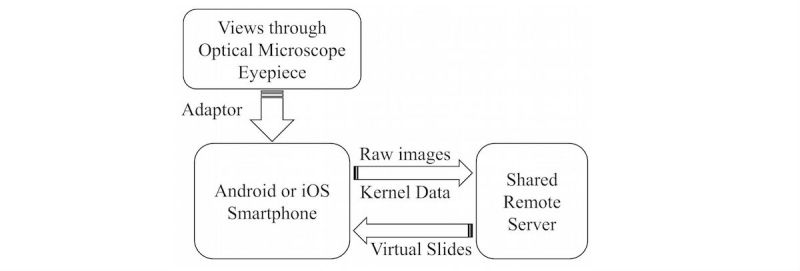
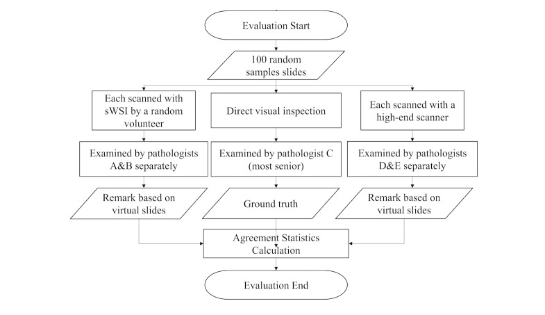
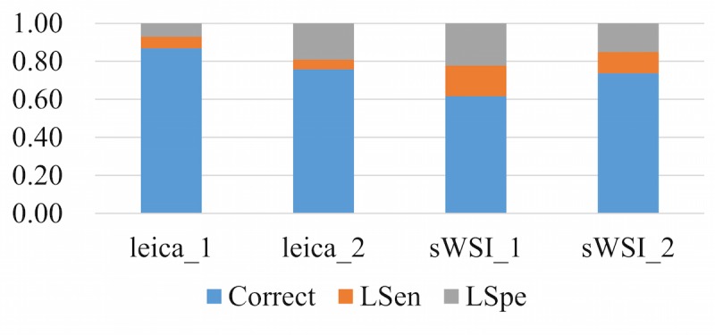
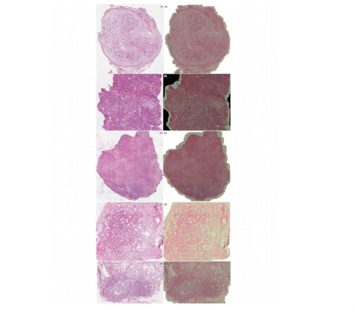
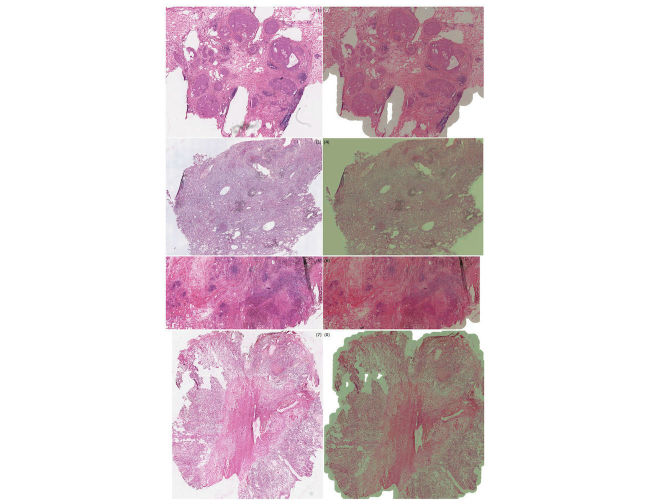
In the sWSI design, the digitization process is split asynchronously between light-weight clients on smartphones and powerful cloud servers. The client apps automatically capture FoVs at up to 12-megapixel resolution and process them in real-time to track the operation of users, then give instant feedback of guidance. The servers first restitch each pair of FoVs, then automatically correct the unknown nonlinear distortion introduced by the lens of the smartphone on the fly, based on pair-wise stitching, before finally combining all FoVs into one gigapixel VS for each scan. These VSs can be viewed using Internet browsers anywhere. In the evaluation experiment, 100 frozen section slides from patients randomly selected among in-patients of the participating hospital were scanned by both a high-end Leica scanner and sWSI. All VSs were examined by senior pathologists whose diagnoses were compared against those made using optical microscopy as ground truth to evaluate the image quality.

RESULTS

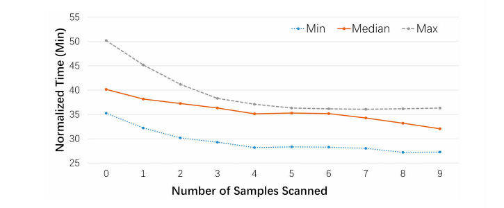
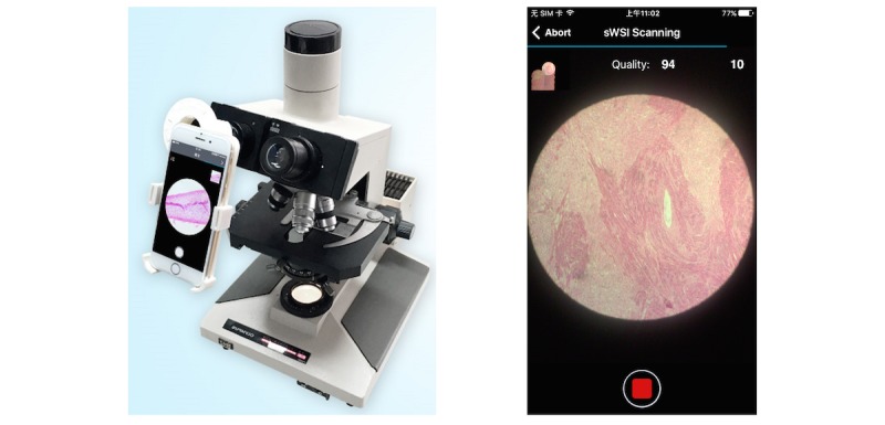
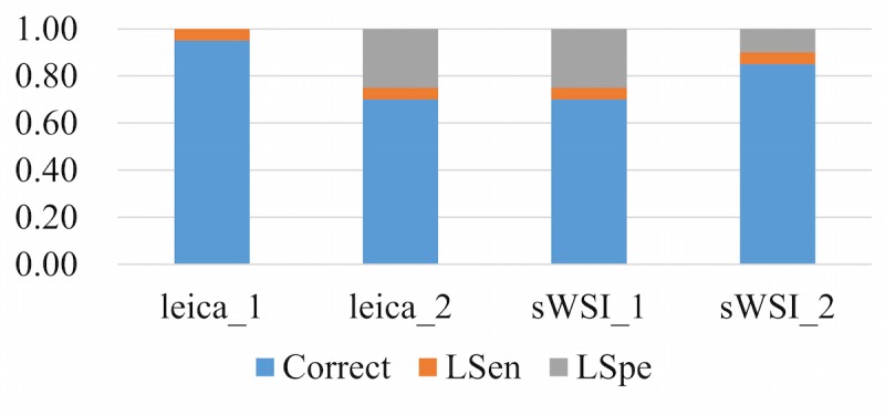
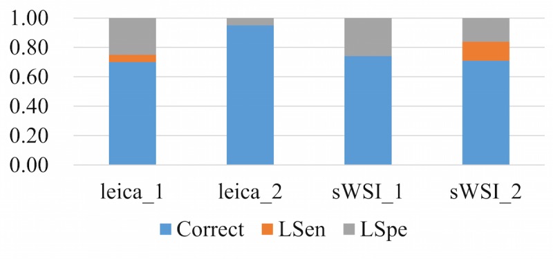
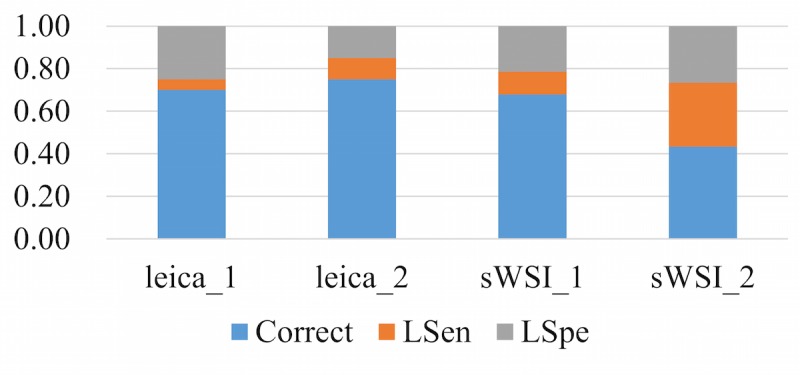
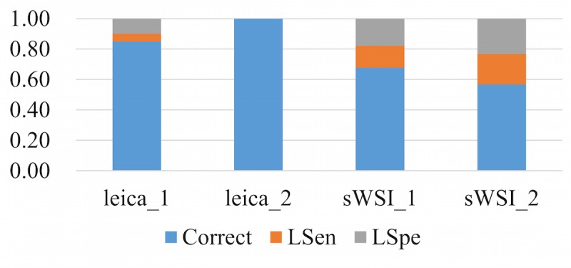
The sWSI system is developed for both Android and iPhone smartphones and is currently being offered to the public. The image quality is reliable and throughput is approximately 1 FoV per second, yielding a 15-by-15 mm slide under 20X object lens in approximately 30-35 minutes, with little training required for the operator. The expected cost for setup is approximately US $100 and scanning each slide costs between US $1 and $10, making sWSI highly cost-effective for infrequent or low-throughput usage. In the clinical evaluation of sample-wise diagnostic reliability, average accuracy scores achieved by sWSI-scan-based diagnoses were as follows: 0.78 for breast, 0.88 for uterine corpus, 0.68 for thyroid, and 0.50 for lung samples. The respective low-sensitivity rates were 0.05, 0.05, 0.13, and 0.25 while the respective low-specificity rates were 0.18, 0.08, 0.20, and 0.25. The participating pathologists agreed that the overall quality of sWSI was generally on par with that produced by high-end scanners, and did not affect diagnosis in most cases. Pathologists confirmed that sWSI is reliable enough for standard diagnoses of most tissue categories, while it can be used for quick screening of difficult cases.

CONCLUSIONS

As an ultra-low-cost alternative to whole slide scanners, diagnosis-ready VS quality and robustness for commercial usage is achieved in the sWSI solution. Operated on main-stream smartphones installed on normal optical microscopes, sWSI readily offers affordable and reliable WSI to resource-limited or infrequent clinical users.

摘要

背景

目标是开发可扩展的全切片成像(sWSI),这是一种基于主流智能手机与常规光学显微镜相结合的全切片成像系统。这种超低成本的解决方案应能提供与独立扫描仪相当的可供诊断的成像质量,支持不同放大倍数的油镜和干镜,并具有合理的高吞吐量。这些性能指标应由专业病理学家进行评估,并与高端扫描仪的指标相匹配。

目的

目标是开发可扩展的全切片成像(sWSI),一种基于智能手机与光学显微镜相结合的全切片成像系统。这种超低成本的解决方案应能提供与独立扫描仪相当的可供诊断的成像质量,支持不同放大倍数的油镜和干镜。所有性能指标应由专业病理学家进行评估,并与高端扫描仪的指标相匹配。

方法

在sWSI设计中,数字化过程在智能手机上的轻量级客户端和强大的云服务器之间异步进行。客户端应用程序自动以高达1200万像素的分辨率捕获视野(FoV),并实时对其进行处理以跟踪用户操作,然后给出即时指导反馈。服务器首先重新拼接每对视野,然后基于成对拼接实时自动校正智能手机镜头引入的未知非线性失真,最后将所有视野组合成每次扫描的一个十亿像素虚拟切片(VS)。这些虚拟切片可以在任何地方使用互联网浏览器查看。在评估实验中,由高端徕卡扫描仪和sWSI对从参与医院的住院患者中随机选择的100例患者的冰冻切片进行扫描。所有虚拟切片均由资深病理学家检查,将其诊断结果与以光学显微镜检查结果作为金标准进行比较,以评估图像质量。

结果

sWSI系统是为安卓和苹果智能手机开发的,目前已向公众提供。图像质量可靠,吞吐量约为每秒1个视野,在20倍物镜下,15×15毫米的切片大约需要30 - 35分钟,操作人员几乎无需培训。预计设置成本约为100美元,扫描每张切片的成本在1美元至10美元之间,这使得sWSI对于不频繁或低吞吐量使用而言具有很高的成本效益。在按样本的诊断可靠性临床评估中,基于sWSI扫描诊断获得的平均准确率如下:乳腺为0.78,子宫体为0.88,甲状腺为0.68,肺样本为0.50。各自的低灵敏度率分别为0.05、0.05、0.13和0.25,各自的低特异性率分别为0.18、0.08、0.20和0.25。参与的病理学家一致认为,sWSI的总体质量通常与高端扫描仪产生的质量相当,并且在大多数情况下不影响诊断。病理学家确认,sWSI对于大多数组织类别的标准诊断足够可靠,同时可用于疑难病例的快速筛查。

结论

作为全切片扫描仪的超低成本替代方案,sWSI解决方案实现了可供诊断的虚拟切片质量和商业使用的稳健性。sWSI安装在普通光学显微镜上,在主流智能手机上运行,可为资源有限或不常使用的临床用户轻松提供经济实惠且可靠的全切片成像。

https://cdn.ncbi.nlm.nih.gov/pmc/blobs/395b/5622289/e65b758682db/mhealth_v5i9e132_fig15.jpg
https://cdn.ncbi.nlm.nih.gov/pmc/blobs/395b/5622289/1604edc2096c/mhealth_v5i9e132_fig1.jpg
https://cdn.ncbi.nlm.nih.gov/pmc/blobs/395b/5622289/6314c4b0f50b/mhealth_v5i9e132_fig2.jpg
https://cdn.ncbi.nlm.nih.gov/pmc/blobs/395b/5622289/4db083430469/mhealth_v5i9e132_fig3.jpg
https://cdn.ncbi.nlm.nih.gov/pmc/blobs/395b/5622289/40350d88a32f/mhealth_v5i9e132_fig4.jpg
https://cdn.ncbi.nlm.nih.gov/pmc/blobs/395b/5622289/b54e2eed37eb/mhealth_v5i9e132_fig5.jpg
https://cdn.ncbi.nlm.nih.gov/pmc/blobs/395b/5622289/eed8fcbf05e3/mhealth_v5i9e132_fig6.jpg
https://cdn.ncbi.nlm.nih.gov/pmc/blobs/395b/5622289/038751c90c30/mhealth_v5i9e132_fig7.jpg
https://cdn.ncbi.nlm.nih.gov/pmc/blobs/395b/5622289/34725e9a914f/mhealth_v5i9e132_fig8.jpg
https://cdn.ncbi.nlm.nih.gov/pmc/blobs/395b/5622289/ac1623787ad5/mhealth_v5i9e132_fig9.jpg
https://cdn.ncbi.nlm.nih.gov/pmc/blobs/395b/5622289/da8e6be03026/mhealth_v5i9e132_fig10.jpg
https://cdn.ncbi.nlm.nih.gov/pmc/blobs/395b/5622289/558f6938a77b/mhealth_v5i9e132_fig11.jpg
https://cdn.ncbi.nlm.nih.gov/pmc/blobs/395b/5622289/0682094f2a7b/mhealth_v5i9e132_fig12.jpg
https://cdn.ncbi.nlm.nih.gov/pmc/blobs/395b/5622289/63b144ef982c/mhealth_v5i9e132_fig13.jpg
https://cdn.ncbi.nlm.nih.gov/pmc/blobs/395b/5622289/069729791561/mhealth_v5i9e132_fig14.jpg
https://cdn.ncbi.nlm.nih.gov/pmc/blobs/395b/5622289/e65b758682db/mhealth_v5i9e132_fig15.jpg

相似文献

1
Development of a Whole Slide Imaging System on Smartphones and Evaluation With Frozen Section Samples.智能手机上全切片成像系统的开发及冷冻切片样本评估
JMIR Mhealth Uhealth. 2017 Sep 15;5(9):e132. doi: 10.2196/mhealth.8242.
2
Development of Whole Slide Imaging on Smartphones and Evaluation With ThinPrep Cytology Test Samples: Follow-Up Study.智能手机上全玻片成像技术的开发及液基薄层细胞学检测样本评估:随访研究
JMIR Mhealth Uhealth. 2018 Apr 4;6(4):e82. doi: 10.2196/mhealth.9518.
3
An array microscope for ultrarapid virtual slide processing and telepathology. Design, fabrication, and validation study.用于超快速虚拟玻片处理和远程病理学的阵列显微镜。设计、制造与验证研究。
Hum Pathol. 2004 Nov;35(11):1303-14. doi: 10.1016/j.humpath.2004.09.002.
4
Evaluation of panoramic digital images using Panoptiq for frozen section diagnosis.使用Panoptiq对全景数字图像进行评估以用于冰冻切片诊断。
J Pathol Inform. 2016 May 4;7:26. doi: 10.4103/2153-3539.181770. eCollection 2016.
5
Diagnostic validation of a portable whole slide imaging scanner for lymphoma diagnosis in resource-constrained setting: A cross-sectional study.资源受限环境下用于淋巴瘤诊断的便携式全切片成像扫描仪的诊断验证:一项横断面研究。
J Pathol Inform. 2023 Jan 9;14:100188. doi: 10.1016/j.jpi.2023.100188. eCollection 2023.
6
Validation of whole slide imaging for frozen section diagnosis of lymph node metastasis: A retrospective study from a tertiary care hospital in Thailand.全切片成像技术在淋巴结转移冰冻切片诊断中的验证:来自泰国一家三级护理医院的回顾性研究。
Ann Diagn Pathol. 2022 Oct;60:151987. doi: 10.1016/j.anndiagpath.2022.151987. Epub 2022 Jun 7.
7
Design of a 3D printed smartphone microscopic system with enhanced imaging ability for biomedical applications.用于生物医学应用的具有增强成像能力的 3D 打印智能手机显微镜系统的设计。
J Microsc. 2019 Oct;276(1):13-20. doi: 10.1111/jmi.12829. Epub 2019 Sep 22.
8
Whole slide imaging (WSI) scanner differences influence optical and computed properties of digitized prostate cancer histology.全切片成像(WSI)扫描仪的差异会影响数字化前列腺癌组织学的光学和计算属性。
J Pathol Inform. 2023 Jul 4;14:100321. doi: 10.1016/j.jpi.2023.100321. eCollection 2023.
9
Validation of a Portable Whole-Slide Imaging System for Frozen Section Diagnosis.用于冰冻切片诊断的便携式全切片成像系统的验证
J Pathol Inform. 2021 Sep 16;12:33. doi: 10.4103/jpi.jpi_95_20. eCollection 2021.
10
Whole slide imaging diagnostic concordance with light microscopy for breast needle biopsies.全玻片成像诊断与乳腺针吸活检的光学显微镜检查一致性。
Hum Pathol. 2014 Aug;45(8):1713-21. doi: 10.1016/j.humpath.2014.04.007. Epub 2014 Apr 24.

引用本文的文献

1
Contemporary Whole Slide Imaging Devices and Their Applications within the Modern Pathology Department: A Selected Hardware Review.当代全玻片成像设备及其在现代病理科的应用:硬件精选综述
J Pathol Inform. 2021 Dec 9;12:50. doi: 10.4103/jpi.jpi_66_21. eCollection 2021.
2
Validation of a Portable Whole-Slide Imaging System for Frozen Section Diagnosis.用于冰冻切片诊断的便携式全切片成像系统的验证
J Pathol Inform. 2021 Sep 16;12:33. doi: 10.4103/jpi.jpi_95_20. eCollection 2021.
3
A Novel Nomogram Based on Machine Learning-Pathomics Signature and Neutrophil to Lymphocyte Ratio for Survival Prediction of Bladder Cancer Patients.

本文引用的文献

1
A Systematic Analysis of Discordant Diagnoses in Digital Pathology Compared With Light Microscopy.与光学显微镜相比,数字病理学中不一致诊断的系统分析。
Arch Pathol Lab Med. 2017 Dec;141(12):1712-1718. doi: 10.5858/arpa.2016-0494-OA. Epub 2017 May 3.
2
Multiview boosting digital pathology analysis of prostate cancer.多视角增强前列腺癌数字病理学分析
Comput Methods Programs Biomed. 2017 Apr;142:91-99. doi: 10.1016/j.cmpb.2017.02.023. Epub 2017 Feb 22.
3
Enterprise Implementation of Digital Pathology: Feasibility, Challenges, and Opportunities.
基于机器学习-病理组学特征和中性粒细胞与淋巴细胞比值的新型列线图用于膀胱癌患者生存预测
Front Oncol. 2021 Jun 17;11:703033. doi: 10.3389/fonc.2021.703033. eCollection 2021.
4
Evaluation and comparison between smartphone and photomicrography based whole slide imaging.基于智能手机和显微摄影的全玻片成像的评估与比较
J Family Med Prim Care. 2020 May 31;9(5):2319-2323. doi: 10.4103/jfmpc.jfmpc_18_20. eCollection 2020 May.
5
Whole Slide Imaging (WSI) in Pathology: Current Perspectives and Future Directions.病理全切片成像(WSI):现状与未来方向。
J Digit Imaging. 2020 Aug;33(4):1034-1040. doi: 10.1007/s10278-020-00351-z.
6
Validation of Remote Digital Frozen Sections for Cancer and Transplant Intraoperative Services.癌症及移植术中服务的远程数字冰冻切片验证
J Pathol Inform. 2018 Oct 9;9:34. doi: 10.4103/jpi.jpi_52_18. eCollection 2018.
7
Long-Range Diagnosis of and Support for Skin Conditions in Field Settings.野外环境中皮肤疾病的远程诊断与支持
Trop Med Infect Dis. 2018 Aug 13;3(3):84. doi: 10.3390/tropicalmed3030084.
8
Development of Whole Slide Imaging on Smartphones and Evaluation With ThinPrep Cytology Test Samples: Follow-Up Study.智能手机上全玻片成像技术的开发及液基薄层细胞学检测样本评估:随访研究
JMIR Mhealth Uhealth. 2018 Apr 4;6(4):e82. doi: 10.2196/mhealth.9518.
企业实施数字病理学:可行性、挑战和机遇。
J Digit Imaging. 2017 Oct;30(5):555-560. doi: 10.1007/s10278-017-9946-9.
4
Digital pathology in clinical use: where are we now and what is holding us back?临床应用中的数字病理学:我们目前的状况以及阻碍我们发展的因素是什么?
Histopathology. 2017 Jan;70(1):134-145. doi: 10.1111/his.12993.
5
Emerging Themes in Image Informatics and Molecular Analysis for Digital Pathology.数字病理学的图像信息学与分子分析中的新兴主题
Annu Rev Biomed Eng. 2016 Jul 11;18:387-412. doi: 10.1146/annurev-bioeng-112415-114722.
6
Digital pathology imaging offers more benefits than glass slides and microscopes.数字病理成像比玻片和显微镜具有更多优势。
BMJ. 2016 Jul 12;354:i3813. doi: 10.1136/bmj.i3813.
7
Image Analysis in Surgical Pathology.外科病理学中的图像分析
Surg Pathol Clin. 2016 Jun;9(2):329-37. doi: 10.1016/j.path.2016.02.001.
8
Evaluation of panoramic digital images using Panoptiq for frozen section diagnosis.使用Panoptiq对全景数字图像进行评估以用于冰冻切片诊断。
J Pathol Inform. 2016 May 4;7:26. doi: 10.4103/2153-3539.181770. eCollection 2016.
9
International Clinical Guidelines for the Adoption of Digital Pathology: A Review of Technical Aspects.数字病理学应用的国际临床指南:技术方面综述
Pathobiology. 2016;83(2-3):99-109. doi: 10.1159/000441192. Epub 2016 Apr 26.
10
New Trends of Emerging Technologies in Digital Pathology.数字病理学中新兴技术的新趋势
Pathobiology. 2016;83(2-3):61-9. doi: 10.1159/000443482. Epub 2016 Apr 26.