Feuerecker Benedikt, Durst Markus, Michalik Michael, Schneider Günter, Saur Dieter, Menzel Marion, Schwaiger Markus, Schilling Franz
Department of Nuclear Medicine, Klinikum rechts der Isar, Technische Universität München, Munich, Germany.
German Cancer Consortium (DKTK) and German Cancer Research Center (DKFZ), Heidelberg, Germany.
J Cancer. 2017 Sep 2;8(15):3078-3085. doi: 10.7150/jca.20250. eCollection 2017.
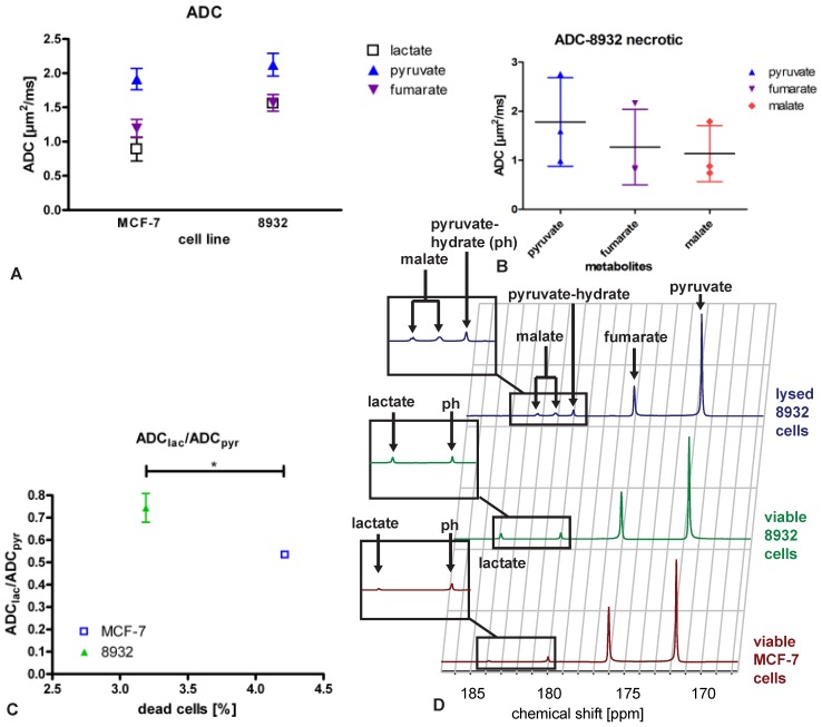
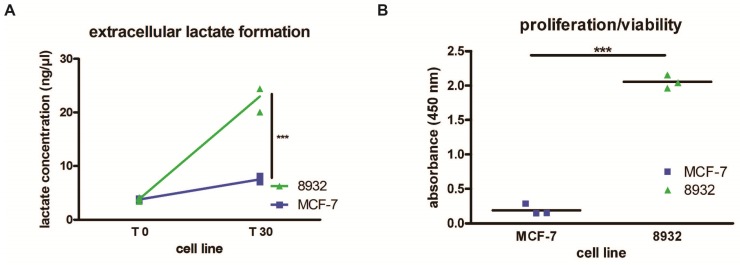
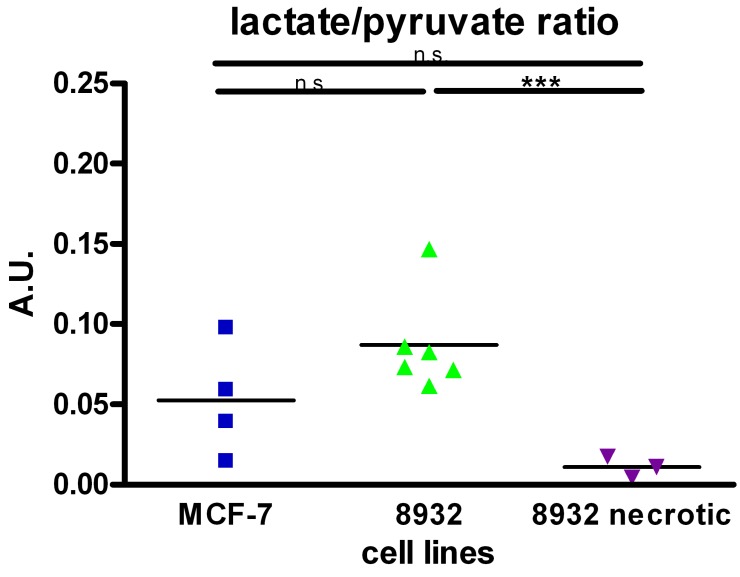
Non-invasive tumor characterization and monitoring are among the key goals of medical imaging. Using hyperpolarized C-labelled metabolic probes fast metabolic pathways can be probed in real-time, providing new opportunities for tumor characterization. In this study, we investigated whether measurement of apparent diffusion coefficient (ADC) measurements and magnetic resonance spectroscopy (MRS) of co-polarized C-labeled pyruvic acid and fumaric acid can non-invasively detect both necrosis and changes in lactate export, which are parameters indicative of tumor aggressiveness. C-labeled pyruvic acid and fumaric acid were co-polarized in a preclinical hyperpolarizer and the dissolved compounds were added to prepared samples of 8932 pancreatic cancer and MCF-7 breast carcinoma cells. Extracellular lactate concentrations and cell viability were measured in separate assays. The mean ratios of the ADC values of lactate and pyruvate (ADC/ADC between MCF-7 (0.533 ± 0.015, n = 3) and 8932 pancreatic cancer cells (0.744 ± 0.064, n = 3) showed a statistically significant difference (p = 0.048). 8932 cells had higher extracellular lactate concentrations in the extracellular medium (22.97 ± 2.53 ng/µl) compared with MCF-7 cells (7.52 ± 0.59 ng/µl; p < 0.001). Fumarate-to-malate conversion was only detectable in necrotic cells, thereby allowing clear differentiation between necrotic and viable cells. We provide evidence that MRS of hyperpolarized C-labelled pyruvic acid and fumaric acid, with their respective conversions to lactate and malate, are useful for characterization of necrosis and lactate efflux in tumor cells.
非侵入性肿瘤特征描述与监测是医学成像的关键目标之一。使用超极化碳标记的代谢探针,可以实时探测快速代谢途径,为肿瘤特征描述提供了新的机会。在本研究中,我们调查了超极化碳标记的丙酮酸和富马酸的表观扩散系数(ADC)测量以及磁共振波谱(MRS)是否能够非侵入性地检测坏死和乳酸输出变化,这两个参数可指示肿瘤侵袭性。碳标记的丙酮酸和富马酸在临床前超极化器中进行超极化,然后将溶解的化合物添加到制备好的8932胰腺癌细胞和MCF-7乳腺癌细胞样本中。在单独的实验中测量细胞外乳酸浓度和细胞活力。MCF-7细胞(0.533±0.015,n = 3)和8932胰腺癌细胞(0.744±0.064,n = 3)之间乳酸和丙酮酸的ADC值平均比值(ADC/ADC)显示出统计学上的显著差异(p = 0.048)。与MCF-7细胞(7.52±0.59 ng/µl;p < 0.001)相比,8932细胞在细胞外培养基中的细胞外乳酸浓度更高(22.97±2.53 ng/µl)。仅在坏死细胞中可检测到富马酸盐向苹果酸盐的转化,从而能够清晰区分坏死细胞和活细胞。我们提供的证据表明,超极化碳标记的丙酮酸和富马酸的MRS及其各自向乳酸和苹果酸的转化,可用于表征肿瘤细胞中的坏死和乳酸流出。