Department of Anatomy, School of Medicine, Tokyo Women's Medical University, 8-1 Kawada-cho, Shinjuku-ku, Tokyo, 162-8666, Japan.
Department of Anatomy, School of Medicine, Toho University, 5-21-16 Omorinishi, Ota-ku, Tokyo, 143-8540, Japan.
Neural Dev. 2017 Sep 20;12(1):17. doi: 10.1186/s13064-017-0094-1.
Cyclin-dependent kinase (CDK) inhibitors play an important role in regulating cell cycle progression, cell cycle exit and cell differentiation. p27 (p27), one of the major CDK inhibitors in the retina, has been shown to control the timing of cell cycle exit of retinal progenitors. However, the precise role of this protein in retinal development remains largely unexplored. We thus analyzed p27-deficient mice to characterize the effects of p27 loss on proliferation, differentiation, and survival of retinal cells.
Expression of p27 in the developing and mature mouse retina was analyzed by immunohistochemistry using antibodies against p27 and cell type-specific markers. Cell proliferation and differentiation were examined in the wild-type and p27-deficient retinas by immunohistochemistry using various cell cycle and differentiation markers.
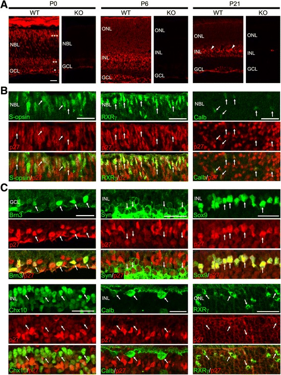
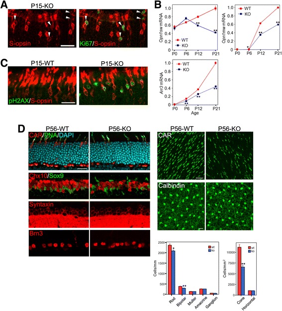
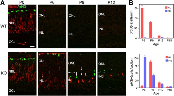
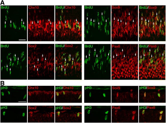
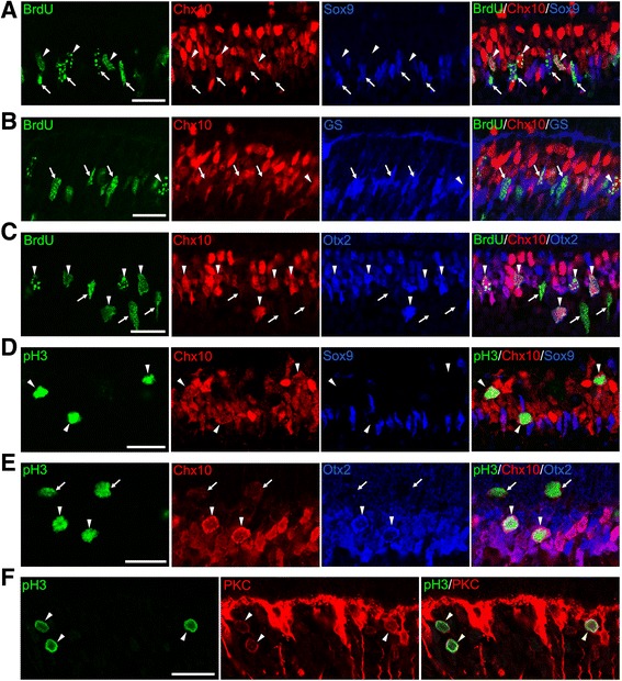
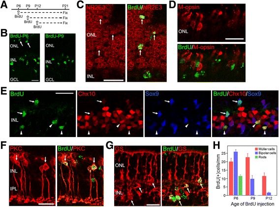
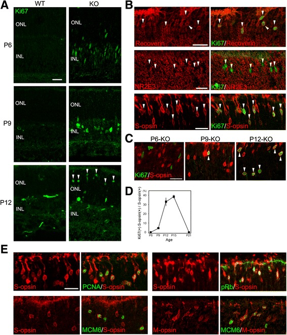
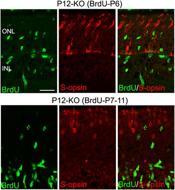
All postmitotic retinal cell types expressed p27 in the mouse retinas. p27 loss caused extension of the period of proliferation in the developing retinas. This extra proliferation was mainly due to ectopic cell cycle reentry of differentiating cells including bipolar cells, Müller glial cells and cones, rather than persistent division of progenitors as previously suggested. Aberrant cell cycle activity of cones was followed by cone death resulting in a significant reduction in cone number in the mature p27-deficient retinas.
Although expressed in all retinal cell types, p27 is required to maintain the quiescence of specific cell types including bipolar cells, Müller glia, and cones while it is dispensable for preventing cell cycle reentry in other cell types.
细胞周期蛋白依赖性激酶 (CDK) 抑制剂在调节细胞周期进程、细胞周期退出和细胞分化方面发挥着重要作用。p27(p27)是视网膜中主要的 CDK 抑制剂之一,已被证明可以控制视网膜祖细胞的细胞周期退出时间。然而,该蛋白在视网膜发育中的精确作用在很大程度上仍未得到探索。因此,我们分析了 p27 缺陷型小鼠,以表征 p27 缺失对视网膜细胞增殖、分化和存活的影响。
使用针对 p27 和细胞类型特异性标志物的抗体通过免疫组织化学分析来分析发育中和成熟的小鼠视网膜中 p27 的表达。通过使用各种细胞周期和分化标志物在野生型和 p27 缺陷型视网膜中检查细胞增殖和分化。
在小鼠视网膜中,所有有丝分裂后视网膜细胞类型都表达 p27。p27 缺失导致发育中视网膜的增殖期延长。这种额外的增殖主要是由于包括双极细胞、Müller 胶质细胞和视锥细胞在内的分化细胞的异位细胞周期重新进入,而不是以前认为的祖细胞持续分裂。视锥细胞异常的细胞周期活动后,视锥细胞死亡导致成熟的 p27 缺陷型视网膜中的视锥细胞数量显著减少。
尽管在所有视网膜细胞类型中表达,但 p27 是维持特定细胞类型(包括双极细胞、Müller 胶质细胞和视锥细胞)静止所必需的,而对于防止其他细胞类型的细胞周期重新进入则是可有可无的。