Völkner Manuela, Kurth Thomas, Schor Jana, Ebner Lynn J A, Bardtke Lara, Kavak Cagri, Hackermüller Jörg, Karl Mike O
German Center for Neurodegenerative Diseases (DZNE) Dresden, Dresden, Germany.
Center for Molecular and Cellular Bioengineering, Technology Platform, Electron Microscopy and Histology Facility, Technische Universität Dresden, Dresden, Germany.
Front Cell Dev Biol. 2021 Apr 27;9:645704. doi: 10.3389/fcell.2021.645704. eCollection 2021.
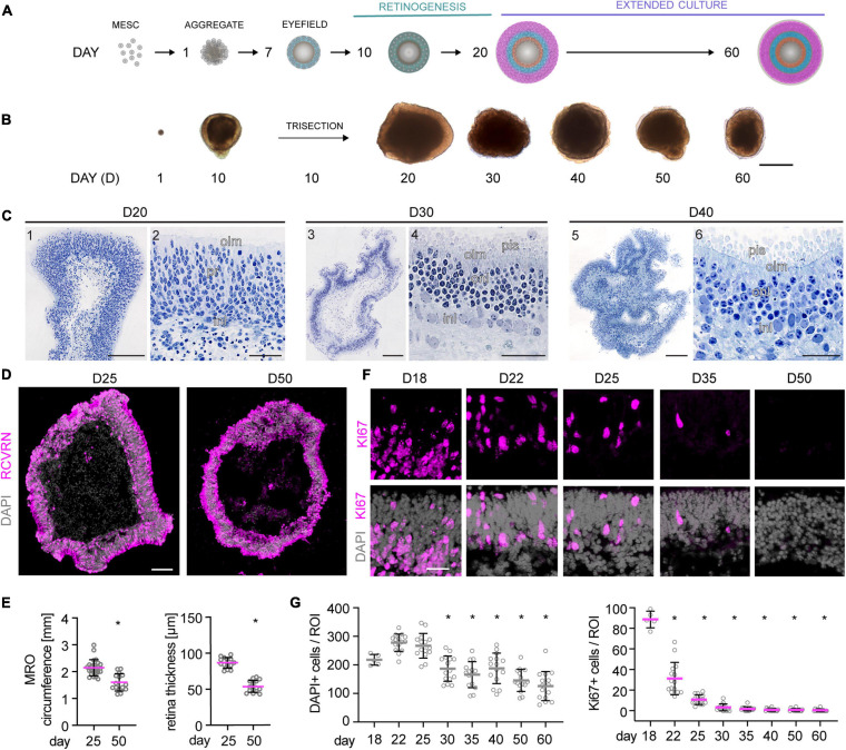
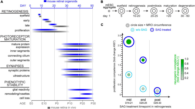
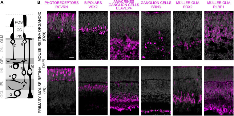
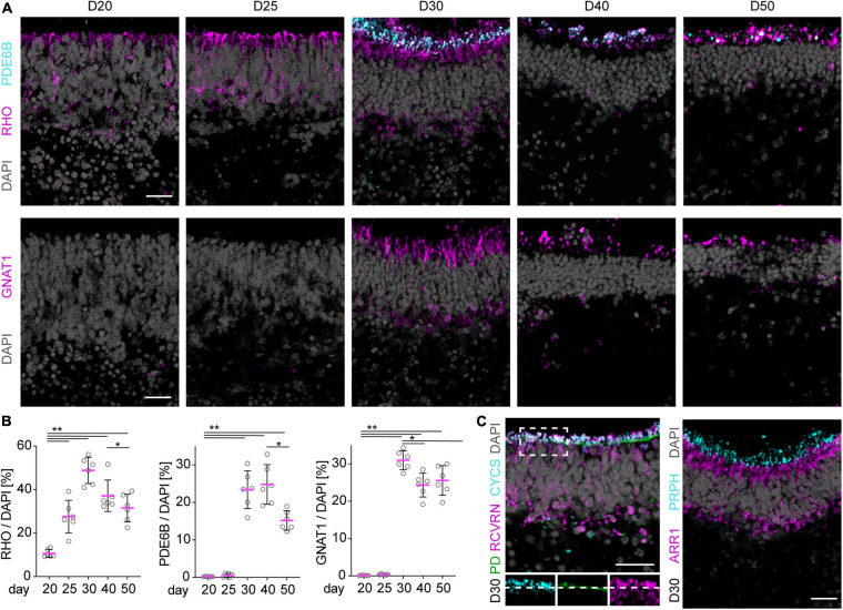
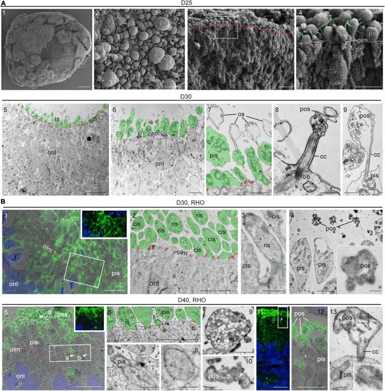
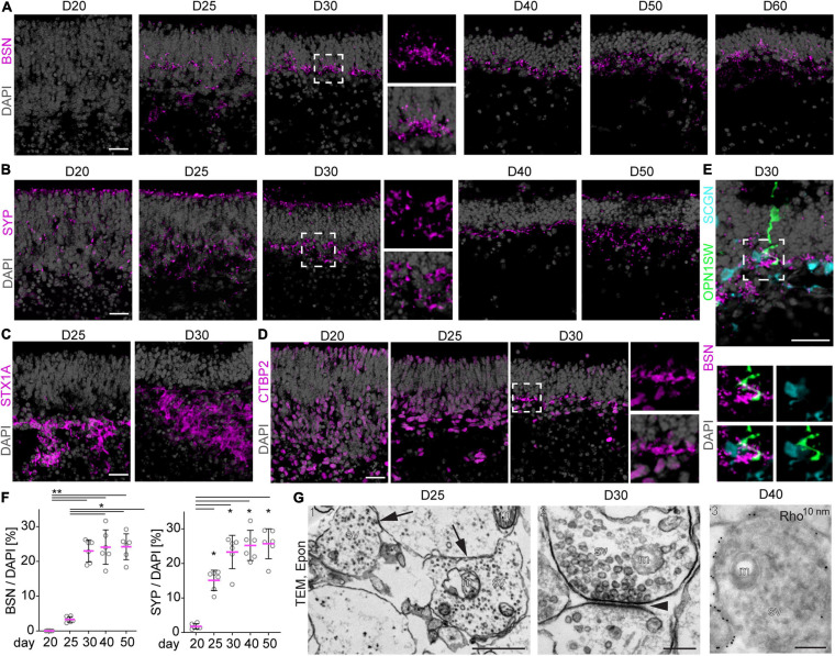
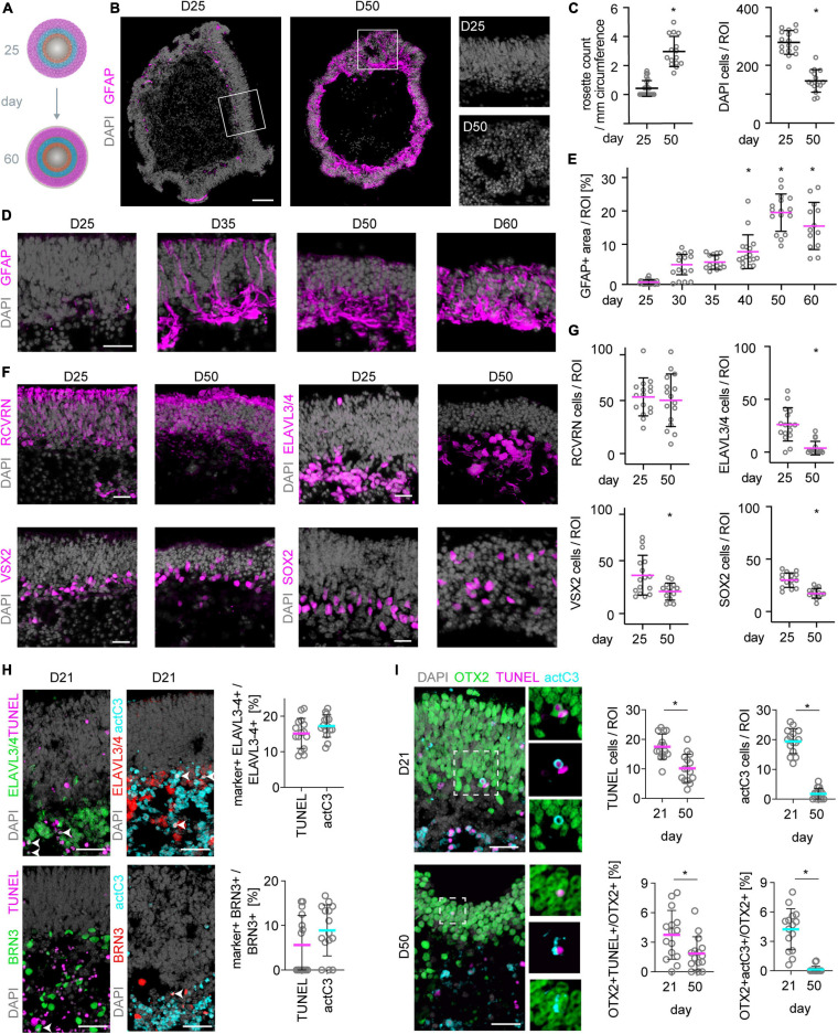
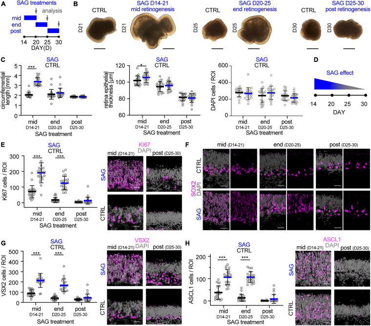
Using retinal organoid systems, organ-like 3D tissues, relies implicitly on their robustness. However, essential key parameters, particularly retinal growth and longer-term culture, are still insufficiently defined. Here, we hypothesize that a previously optimized protocol for high yield of evenly-sized mouse retinal organoids with low variability facilitates assessment of such parameters. We demonstrate that these organoids reliably complete retinogenesis, and can be maintained at least up to 60 days in culture. During this time, the organoids continue to mature on a molecular and (ultra)structural level: They develop photoreceptor outer segments and synapses, transiently maintain its cell composition for about 5-10 days after completing retinogenesis, and subsequently develop pathologic changes - mainly of the inner but also outer retina and reactive gliosis. To test whether this organoid system provides experimental access to the retina during and upon completion of development, we defined and stimulated organoid growth by activating sonic hedgehog signaling, which in patients and mice with a congenital defect leads to enlarged eyes. Here, a sonic hedgehog signaling activator increased retinal epithelia length in the organoid system when applied during but not after completion of development. This experimentally supports organoid maturation, stability, and experimental reproducibility in this organoid system, and provides a potential enlarged retina pathology model, as well as a protocol for producing larger organoids. Together, our study advances the understanding of retinal growth, maturation, and maintenance, and further optimizes the organoid system for future utilization.
利用视网膜类器官系统,即类器官样三维组织,隐含地依赖于它们的稳健性。然而,关键参数,尤其是视网膜生长和长期培养,仍未得到充分界定。在此,我们假设,一种先前优化的方案,用于高产、大小均匀且变异性低的小鼠视网膜类器官,有助于评估此类参数。我们证明,这些类器官能可靠地完成视网膜发育,并且在培养中至少可维持60天。在此期间,类器官在分子和(超)结构水平上持续成熟:它们发育出光感受器外段和突触,在完成视网膜发育后短暂维持其细胞组成约5 - 10天,随后出现病理变化——主要在内层视网膜,但外层视网膜和反应性胶质增生也有变化。为了测试这个类器官系统在发育期间及发育完成后是否能提供对视网膜的实验性研究途径,我们通过激活音猬因子信号通路来定义和刺激类器官生长,在患有先天性缺陷的患者和小鼠中,该信号通路会导致眼睛增大。在此,当在发育期间而非发育完成后应用时,音猬因子信号通路激活剂增加了类器官系统中视网膜上皮的长度。这在实验上支持了该类器官系统中的类器官成熟、稳定性和实验可重复性,并提供了一个潜在的扩大视网膜病理模型,以及一种生产更大类器官的方案。总之,我们的研究推进了对视网膜生长、成熟和维持的理解,并进一步优化了类器官系统以供未来使用。