• 文献检索
  • 文档翻译
  • 深度研究
  • 学术资讯
  • Suppr Zotero 插件Zotero 插件
  • 邀请有礼
  • 套餐&价格
  • 历史记录
应用&插件
Suppr Zotero 插件Zotero 插件浏览器插件Mac 客户端Windows 客户端微信小程序
定价
高级版会员购买积分包购买API积分包
服务
文献检索文档翻译深度研究API 文档MCP 服务
关于我们
关于 Suppr公司介绍联系我们用户协议隐私条款
关注我们

Suppr 超能文献

核心技术专利:CN118964589B侵权必究
粤ICP备2023148730 号-1Suppr @ 2026

文献检索

告别复杂PubMed语法,用中文像聊天一样搜索,搜遍4000万医学文献。AI智能推荐,让科研检索更轻松。

立即免费搜索

文件翻译

保留排版,准确专业,支持PDF/Word/PPT等文件格式,支持 12+语言互译。

免费翻译文档

深度研究

AI帮你快速写综述,25分钟生成高质量综述,智能提取关键信息,辅助科研写作。

立即免费体验

福尔马林固定和冷冻切片仅导致眼组织的形状或大小发生最小变化。

Formalin Fixation and Cryosectioning Cause Only Minimal Changes in Shape or Size of Ocular Tissues.

机构信息

Department of Ophthalmology, University of Pittsburgh, Pittsburgh, PA, 15213, USA.

Department of Bioengineering, University of Pittsburgh, Pittsburgh, PA, 15213, USA.

出版信息

Sci Rep. 2017 Sep 21;7(1):12065. doi: 10.1038/s41598-017-12006-1.

DOI:10.1038/s41598-017-12006-1
PMID:28935889
原文链接:https://pmc.ncbi.nlm.nih.gov/articles/PMC5608899/
Abstract

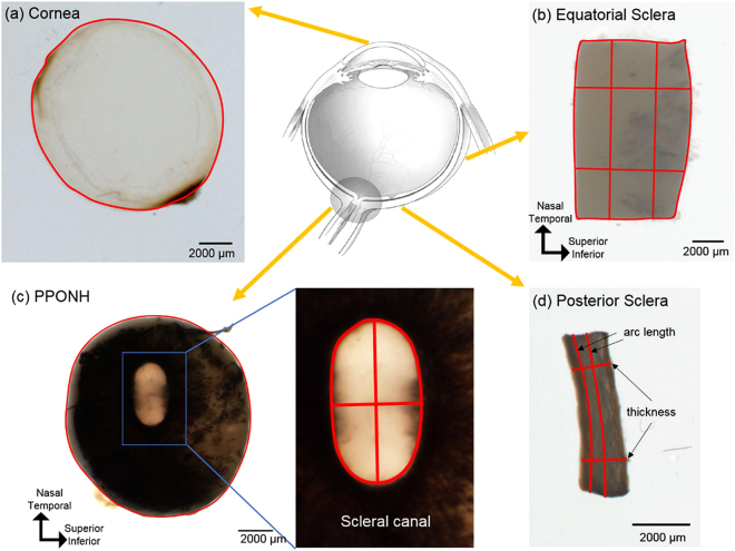
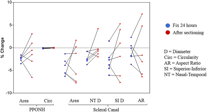
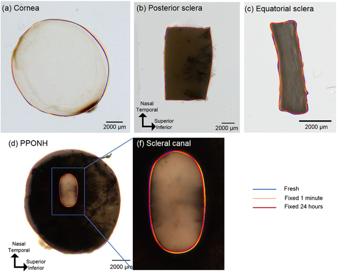
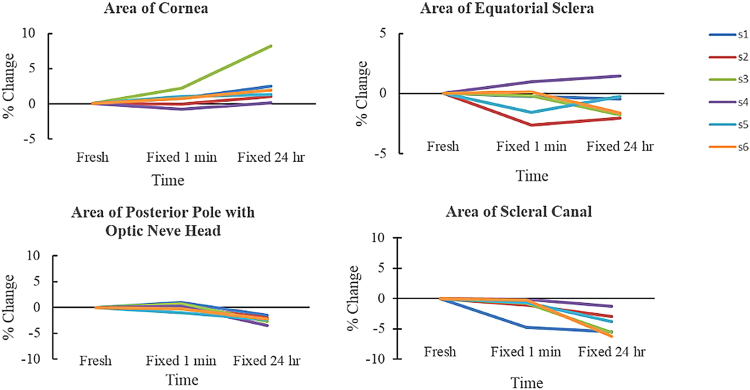
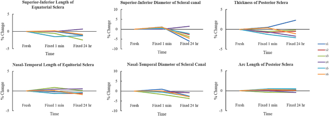
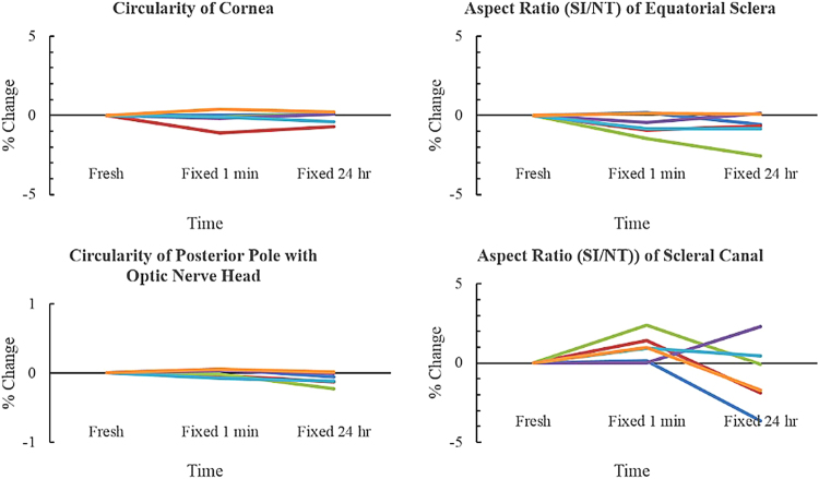
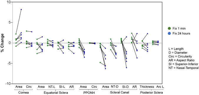
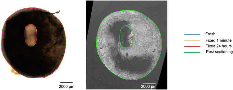
Advances in imaging have made it increasingly common to study soft tissues without first embedding them in plastic or paraffin and without using labels or stains. The process, however, usually still involves fixation and cryosectioning, which could deform the tissues. Our goal was to quantify the morphological changes of ocular tissues caused by formalin fixation and cryosectioning. From each of 6 porcine eyes, 4 regions were obtained: cornea, equatorial and posterior sclera, and posterior pole containing the optic nerve head. Samples were imaged using visible light microscopy fresh, 1-minute and 24-hours post-fixation, and post-cryosectioning. Effects were assessed by 14 parameters representing sample size and shape. Overall, formalin fixation and sectioning caused only minimal changes to the ocular tissues, with average percentage parameter differences of 0.1%, 1%, and 1.2% between fresh and post-fixing by 1 minute, 24 hours, and post-cryosectioning, respectively. Parameter changes were not directional, and were only weakly dependent on the duration of fixation and the region of the eye. These results demonstrate that formalin fixation and cryosectioning are good choices for studying ocular tissue morphology and structure, as they do not cause the large tissue shrinkage or distortions typically associated with other, more complicated, techniques.

摘要

成像技术的进步使得人们越来越倾向于在不首先将软组织嵌入塑料或石蜡中、不使用标记物或染色剂的情况下对其进行研究。然而,该过程通常仍然涉及固定和冷冻切片,这可能会导致组织变形。我们的目标是量化甲醛固定和冷冻切片对眼部组织形态的变化。从 6 只猪眼的每只眼中获得 4 个区域:角膜、赤道和后巩膜以及包含视神经头的后极。新鲜、固定后 1 分钟和 24 小时以及冷冻切片后,使用可见光显微镜对样本进行成像。通过代表样本大小和形状的 14 个参数评估效果。总体而言,甲醛固定和切片仅对眼部组织造成最小的变化,新鲜和固定后 1 分钟、24 小时以及冷冻切片后分别有 0.1%、1%和 1.2%的平均百分比参数差异。参数变化没有方向性,仅与固定时间和眼部区域的持续时间有弱相关性。这些结果表明,甲醛固定和冷冻切片是研究眼部组织形态和结构的良好选择,因为它们不会导致与其他更复杂技术相关的大组织收缩或变形。

https://cdn.ncbi.nlm.nih.gov/pmc/blobs/e2e4/5608899/14bf329ab91a/41598_2017_12006_Fig8_HTML.jpg
https://cdn.ncbi.nlm.nih.gov/pmc/blobs/e2e4/5608899/a78f09d7c403/41598_2017_12006_Fig1_HTML.jpg
https://cdn.ncbi.nlm.nih.gov/pmc/blobs/e2e4/5608899/706f4d443a2b/41598_2017_12006_Fig2_HTML.jpg
https://cdn.ncbi.nlm.nih.gov/pmc/blobs/e2e4/5608899/bed74428ab87/41598_2017_12006_Fig3_HTML.jpg
https://cdn.ncbi.nlm.nih.gov/pmc/blobs/e2e4/5608899/c39633eaae97/41598_2017_12006_Fig4_HTML.jpg
https://cdn.ncbi.nlm.nih.gov/pmc/blobs/e2e4/5608899/52e3e1ea02b6/41598_2017_12006_Fig5_HTML.jpg
https://cdn.ncbi.nlm.nih.gov/pmc/blobs/e2e4/5608899/0de38d6d3861/41598_2017_12006_Fig6_HTML.jpg
https://cdn.ncbi.nlm.nih.gov/pmc/blobs/e2e4/5608899/be94f4fbc717/41598_2017_12006_Fig7_HTML.jpg
https://cdn.ncbi.nlm.nih.gov/pmc/blobs/e2e4/5608899/14bf329ab91a/41598_2017_12006_Fig8_HTML.jpg

相似文献

1
Formalin Fixation and Cryosectioning Cause Only Minimal Changes in Shape or Size of Ocular Tissues.福尔马林固定和冷冻切片仅导致眼组织的形状或大小发生最小变化。
Sci Rep. 2017 Sep 21;7(1):12065. doi: 10.1038/s41598-017-12006-1.
2
Cryostat sectioning of formalin-fixed brain: further attempt to improve section quality by previous infiltration with O.C.T. compound.福尔马林固定脑的低温恒冷箱切片:通过预先用O.C.T. 复合物浸润进一步尝试提高切片质量。
Tohoku J Exp Med. 1993 Oct;171(2):101-5. doi: 10.1620/tjem.171.101.
3
Cryosectioning.冷冻切片术
Methods Mol Biol. 2013;1018:301-11. doi: 10.1007/978-1-62703-444-9_28.
4
Fresh Frozen Versus Formalin-Fixed Paraffin Embedded for Mass Spectrometry Imaging.用于质谱成像的新鲜冷冻样本与福尔马林固定石蜡包埋样本的比较
Methods Mol Biol. 2017;1618:7-14. doi: 10.1007/978-1-4939-7051-3_2.
5
Laser Microdissection-Based Microproteomics of Formalin-Fixed and Paraffin-Embedded (FFPE) Tissues.基于激光显微切割技术的福尔马林固定石蜡包埋(FFPE)组织的微蛋白质组学
Methods Mol Biol. 2018;1723:19-31. doi: 10.1007/978-1-4939-7558-7_2.
6
Cryosectioning.冷冻切片术
Curr Protoc Pharmacol. 2001 May;Appendix 3:3E. doi: 10.1002/0471141755.pha03es07.
7
The use of commercially available adhesive tapes to preserve cartilage and bone tissue integrity during cryosectioning.在冷冻切片过程中使用市售胶带以保持软骨和骨组织的完整性。
Biotechniques. 2018 Oct;65(4):191-196. doi: 10.2144/btn-2018-0021.
8
An alternative means of retaining ocular structure and improving immunoreactivity for light microscopy studies.一种用于保留眼部结构并提高免疫反应性以进行光学显微镜研究的替代方法。
Mol Vis. 2015 Apr 16;21:428-42. eCollection 2015.
9
Will PAXgene substitute formalin? A morphological and molecular comparative study using a new fixative system.PAXgene 是否会替代福尔马林?一种新固定系统的形态学和分子比较研究。
J Clin Pathol. 2013 Feb;66(2):124-35. doi: 10.1136/jclinpath-2012-200983. Epub 2012 Nov 3.
10
Biomedical analysis of formalin-fixed, paraffin-embedded tissue samples: The Holy Grail for molecular diagnostics.福尔马林固定、石蜡包埋组织样本的生物医学分析:分子诊断的圣杯。
J Pharm Biomed Anal. 2018 Jun 5;155:125-134. doi: 10.1016/j.jpba.2018.03.065. Epub 2018 Apr 2.

引用本文的文献

1
Multiscale analysis of equatorial sclera anisotropy: Revealing discrepancies in fiber orientation and mechanical properties.赤道部巩膜各向异性的多尺度分析:揭示纤维取向和力学性能的差异
Sci Adv. 2025 Jul 11;11(28):eadp8631. doi: 10.1126/sciadv.adp8631. Epub 2025 Jul 9.
2
Humpback whale () visual acuity allows silhouette detection but not fine detail discrimination over ecological distances.座头鲸的视力能够探测到轮廓,但在生态距离范围内无法辨别精细细节。
Proc Biol Sci. 2025 May;292(2047):20243101. doi: 10.1098/rspb.2024.3101. Epub 2025 May 21.
3
Impact of Elevated Intraocular Pressure on Lamina Cribrosa Oxygenation: A Combined Experimental-Computational Study on Monkeys.

本文引用的文献

1
Collagen Architecture of the Posterior Pole: High-Resolution Wide Field of View Visualization and Analysis Using Polarized Light Microscopy.后极部的胶原结构:使用偏振光显微镜的高分辨率宽视野可视化与分析
Invest Ophthalmol Vis Sci. 2017 Feb 1;58(2):735-744. doi: 10.1167/iovs.16-20772.
2
Polarization microscopy for characterizing fiber orientation of ocular tissues.用于表征眼组织纤维取向的偏振显微镜。
Biomed Opt Express. 2015 Nov 5;6(12):4705-18. doi: 10.1364/BOE.6.004705. eCollection 2015 Dec 1.
3
Correcting the Shrinkage Effects of Formalin Fixation and Tissue Processing for Renal Tumors: toward Standardization of Pathological Reporting of Tumor Size.
眼压升高对筛板氧合的影响:一项对猴子的实验与计算相结合的研究
Ophthalmol Sci. 2025 Jan 31;5(3):100725. doi: 10.1016/j.xops.2025.100725. eCollection 2025 May-Jun.
4
Anatomical assessment of the Kambin's triangle for percutaneous posterolateral transforaminal endoscopic surgery of lumbar intervertebral discs: a magnetic resonance imaging based study.基于磁共振成像的经皮后外侧椎间孔镜下腰椎间盘手术中Kambin三角的解剖学评估研究
Anat Cell Biol. 2024 Dec 31;57(4):523-534. doi: 10.5115/acb.24.112. Epub 2024 Nov 8.
5
Proposing a Methodology for Axon-Centric Analysis of IOP-Induced Mechanical Insult.提出一种针对眼压诱导的机械损伤的轴突中心分析方法。
Invest Ophthalmol Vis Sci. 2024 Nov 4;65(13):1. doi: 10.1167/iovs.65.13.1.
6
Capturing sclera anisotropy using direct collagen fiber models. Linking microstructure to macroscopic mechanical properties.使用直接胶原纤维模型捕捉巩膜各向异性。将微观结构与宏观力学性能联系起来。
bioRxiv. 2024 Sep 27:2024.09.12.612702. doi: 10.1101/2024.09.12.612702.
7
Impact of elevated IOP on lamina cribrosa oxygenation; A combined experimental-computational study on monkeys.眼压升高对筛板氧合的影响;一项针对猴子的实验与计算相结合的研究
bioRxiv. 2024 Sep 10:2024.09.05.609208. doi: 10.1101/2024.09.05.609208.
8
Impact of anatomic variability and other vascular factors on lamina cribrosa hypoxia.解剖变异及其他血管因素对筛板缺氧的影响。
bioRxiv. 2024 Sep 13:2024.09.12.610282. doi: 10.1101/2024.09.12.610282.
9
A method for analyzing AFM force mapping data obtained from soft tissue cryosections.一种分析从软组织冷冻切片获得的原子力显微镜力映射数据的方法。
J Biomech. 2024 May;168:112113. doi: 10.1016/j.jbiomech.2024.112113. Epub 2024 Apr 19.
10
Lamina Cribrosa Insertions Into the Sclera Are Sparser, Narrower, and More Slanted in the Anterior Lamina.前层视乳头筛板插入巩膜处的板层插入物更稀疏、更狭窄且更倾斜。
Invest Ophthalmol Vis Sci. 2024 Apr 1;65(4):35. doi: 10.1167/iovs.65.4.35.
纠正福尔马林固定和组织处理对肾肿瘤的收缩效应:实现肿瘤大小病理报告标准化。
J Cancer. 2015 Jul 2;6(8):759-66. doi: 10.7150/jca.12094. eCollection 2015.
4
Quantitative analysis of three-dimensional fibrillar collagen microstructure within the normal, aged and glaucomatous human optic nerve head.正常、老龄和青光眼患者视神经头内三维纤维状胶原微观结构的定量分析
J R Soc Interface. 2015 May 6;12(106). doi: 10.1098/rsif.2015.0066.
5
Residual deformations in ocular tissues.眼部组织中的残余变形。
J R Soc Interface. 2015 Apr 6;12(105). doi: 10.1098/rsif.2014.1101.
6
Quantitative mapping of collagen fiber orientation in non-glaucoma and glaucoma posterior human sclerae.定量绘制非青光眼和青光眼人后巩膜胶原纤维方向图。
Invest Ophthalmol Vis Sci. 2012 Aug 7;53(9):5258-70. doi: 10.1167/iovs.12-9705.
7
Fiji: an open-source platform for biological-image analysis.斐济:一个用于生物影像分析的开源平台。
Nat Methods. 2012 Jun 28;9(7):676-82. doi: 10.1038/nmeth.2019.
8
Shrinkage revisited: how long is long enough?再谈回缩:多长时间才算足够长?
Ophthalmic Plast Reconstr Surg. 2012 Jul-Aug;28(4):261-3. doi: 10.1097/IOP.0b013e31824ee720.
9
Quantitative mapping of scleral fiber orientation in normal rat eyes.正常大鼠眼球巩膜纤维方向的定量测绘。
Invest Ophthalmol Vis Sci. 2011 Dec 28;52(13):9684-93. doi: 10.1167/iovs.11-7894.
10
Dimensions of the human sclera: Thickness measurement and regional changes with axial length.人眼球巩膜的维度:厚度测量及其与眼轴长度的区域变化。
Exp Eye Res. 2010 Feb;90(2):277-84. doi: 10.1016/j.exer.2009.11.001. Epub 2009 Nov 11.