• 文献检索
  • 文档翻译
  • 深度研究
  • 学术资讯
  • Suppr Zotero 插件Zotero 插件
  • 邀请有礼
  • 套餐&价格
  • 历史记录
应用&插件
Suppr Zotero 插件Zotero 插件浏览器插件Mac 客户端Windows 客户端微信小程序
定价
高级版会员购买积分包购买API积分包
服务
文献检索文档翻译深度研究API 文档MCP 服务
关于我们
关于 Suppr公司介绍联系我们用户协议隐私条款
关注我们

Suppr 超能文献

核心技术专利:CN118964589B侵权必究
粤ICP备2023148730 号-1Suppr @ 2026

文献检索

告别复杂PubMed语法,用中文像聊天一样搜索,搜遍4000万医学文献。AI智能推荐,让科研检索更轻松。

立即免费搜索

文件翻译

保留排版,准确专业,支持PDF/Word/PPT等文件格式,支持 12+语言互译。

免费翻译文档

深度研究

AI帮你快速写综述,25分钟生成高质量综述,智能提取关键信息,辅助科研写作。

立即免费体验

肿瘤坏死因子诱导基因 6 蛋白通过促进小鼠自噬形成来改善慢性肝损伤。

Tumor necrosis factor-inducible gene 6 protein ameliorates chronic liver damage by promoting autophagy formation in mice.

机构信息

Department of Integrated Biological Science, College of Natural Science, Pusan National University, Pusan, Korea.

Department of Life Science, Sogang University, Seoul, Korea.

出版信息

Exp Mol Med. 2017 Sep 22;49(9):e380. doi: 10.1038/emm.2017.140.

DOI:10.1038/emm.2017.140
PMID:28935975
原文链接:https://pmc.ncbi.nlm.nih.gov/articles/PMC5628276/
Abstract

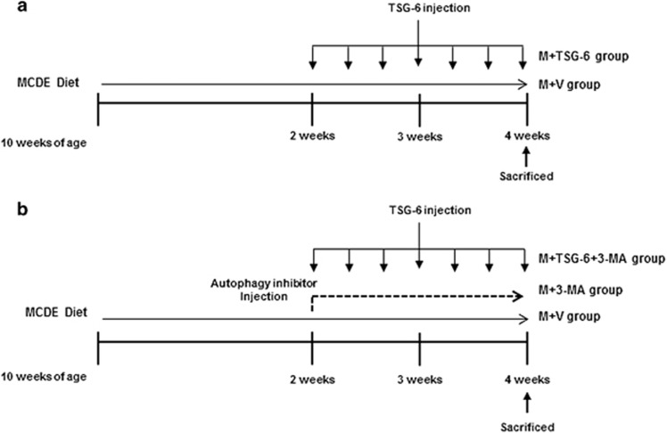
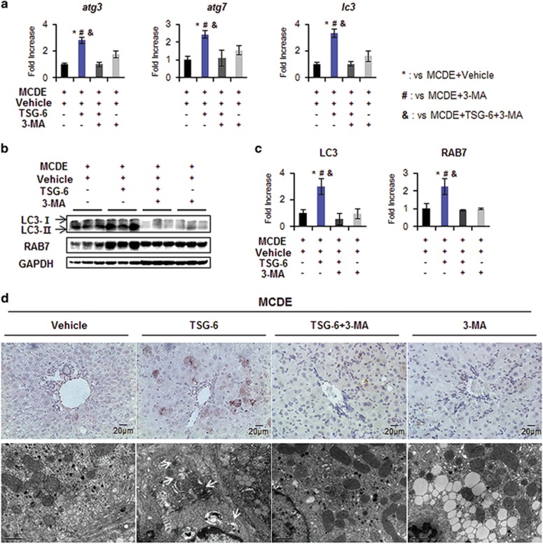
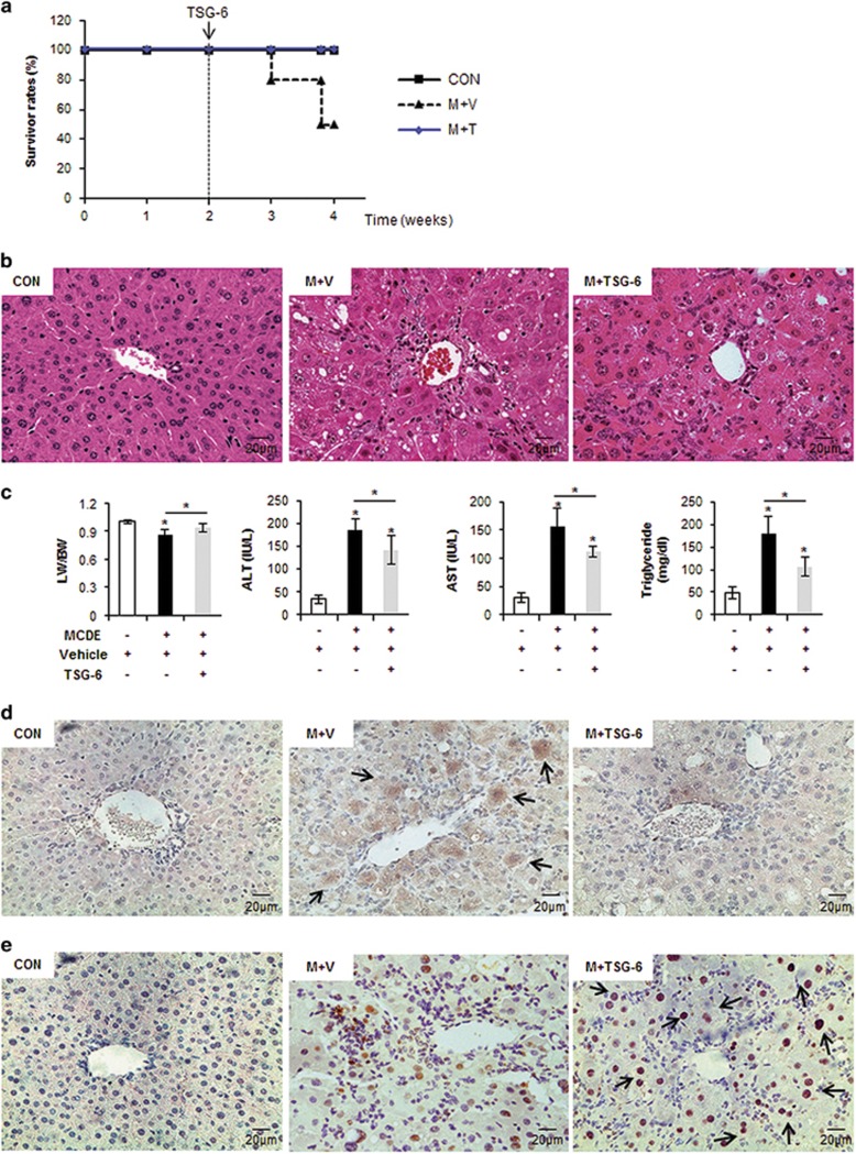
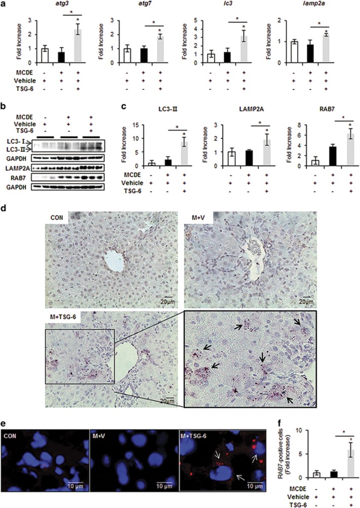
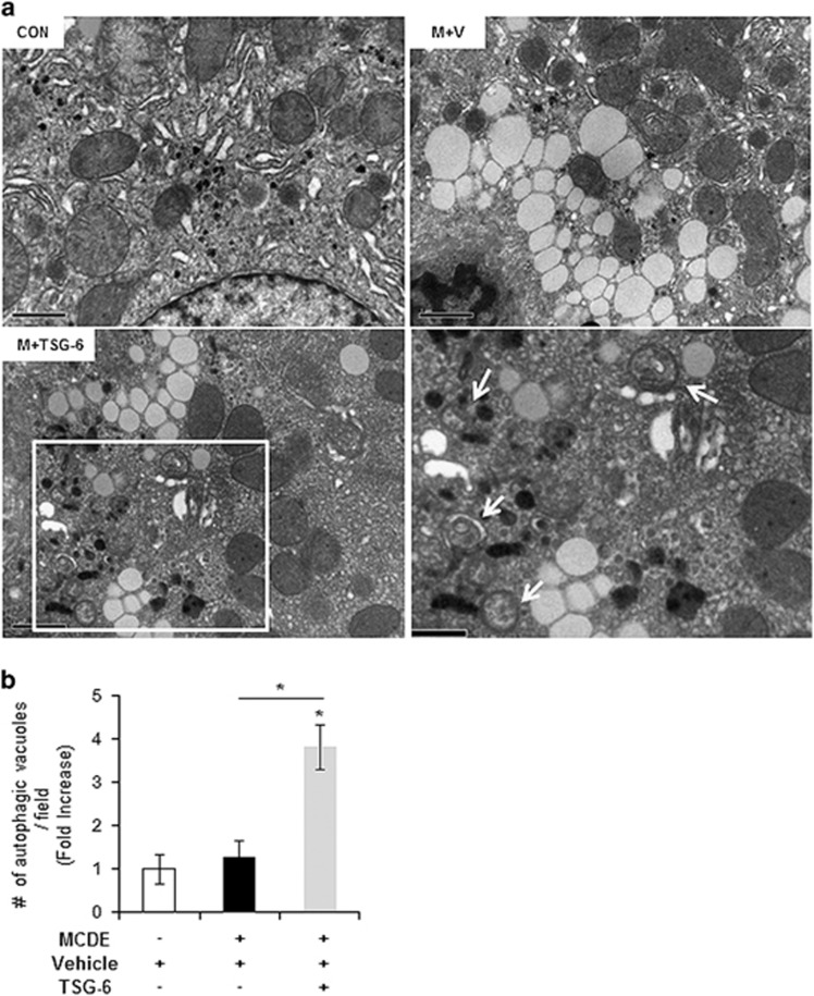
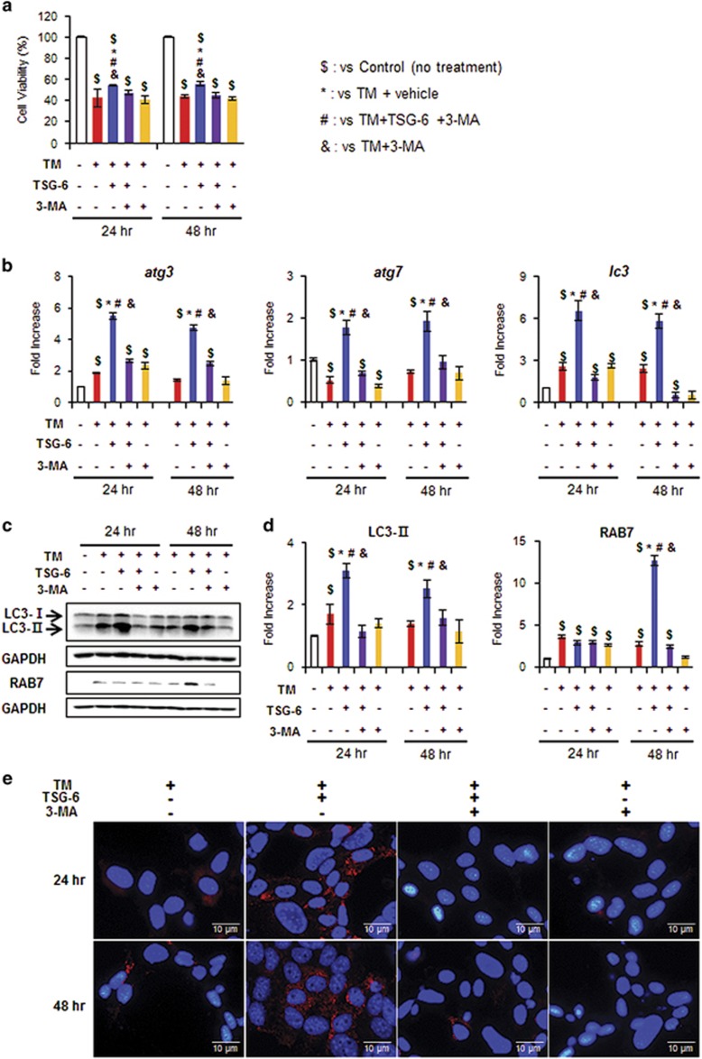
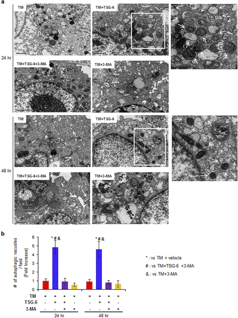
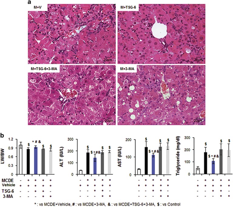
Tumor necrosis factor-inducible gene 6 protein (TSG-6) has recently been shown to protect the liver from acute damage. However, the mechanism underlying the effect of TSG-6 on the liver remains unclear. Autophagy is a catabolic process that targets cell components to lysosomes for degradation, and its functions are reported to be dysregulated in liver diseases. Here we investigate whether TSG-6 promotes liver regeneration by inducing autophagic clearance in damaged livers. Mice fed a methionine choline-deficient diet supplemented with 0.1% ethionine (MCDE) for 2 weeks were injected with TSG-6 (the M+TSG-6 group) or saline (the M+V group) and fed with MCDE for 2 additional weeks. Histomorphological evidence of injury and increased levels of liver enzymes were evident in MCDE-treated mice, whereas these symptoms were ameliorated in the M+TSG-6 group. Livers from this group contained less active caspase-3 and more Ki67-positive hepatocytic cells than the M+V group. The autophagy markers ATG3, ATG7, LC3-II, LAMP2A and RAB7 were elevated in the M+TSG-6 group compared with those in the M+V group. Immunostaining for LC3 and RAB7 and electron microscopy analysis showed the accumulation of autophagy structures in the M+TSG-6 group. TSG-6 also blocked both tunicamycin- and palmitate-induced apoptosis of hepatocytes and increased their viability by inducing autophagy formation in these cells. An autophagy inhibitor suppressed TSG-6-mediated autophagy in the injured hepatocytes and livers of MCDE-treated mice. These results therefore demonstrate that TSG-6 protects hepatocytes from damage by enhancing autophagy influx and contributes to liver regeneration, suggesting that TSG-6 has therapeutic potential for the treatment of liver diseases.

摘要

肿瘤坏死因子诱导基因 6 蛋白(TSG-6)最近被证明可以保护肝脏免受急性损伤。然而,TSG-6 对肝脏影响的机制尚不清楚。自噬是一种靶向溶酶体进行降解的细胞成分的分解代谢过程,其功能在肝脏疾病中被报道失调。在这里,我们研究了 TSG-6 是否通过诱导受损肝脏中的自噬清除来促进肝脏再生。用蛋氨酸胆碱缺乏饲料补充 0.1%乙硫氨酸(MCDE)喂养 2 周的小鼠注射 TSG-6(M+TSG-6 组)或生理盐水(M+V 组),并再用 MCDE 喂养 2 周。MCDE 处理的小鼠有明显的损伤组织形态学证据和肝酶水平升高,而 M+TSG-6 组的这些症状得到了改善。与 M+V 组相比,该组的肝脏中活性 caspase-3 较少,Ki67 阳性肝细胞较多。与 M+V 组相比,M+TSG-6 组的自噬标记物 ATG3、ATG7、LC3-II、LAMP2A 和 RAB7 升高。免疫组化 LC3 和 RAB7 以及电子显微镜分析显示,M+TSG-6 组自噬结构积累。TSG-6 还通过诱导这些细胞中自噬形成,阻断了衣霉素和棕榈酸诱导的肝细胞凋亡,并提高了它们的活力。自噬抑制剂抑制了 TSG-6 在 MCDE 处理的小鼠受损肝细胞和肝脏中的自噬作用。这些结果表明,TSG-6 通过增强自噬流入来保护肝细胞免受损伤,并有助于肝脏再生,表明 TSG-6 具有治疗肝脏疾病的潜力。

https://cdn.ncbi.nlm.nih.gov/pmc/blobs/d4a5/5628276/cbf301b6ac24/emm2017140f8.jpg
https://cdn.ncbi.nlm.nih.gov/pmc/blobs/d4a5/5628276/b7d07a4bcc4c/emm2017140f1.jpg
https://cdn.ncbi.nlm.nih.gov/pmc/blobs/d4a5/5628276/5fa32272ee6d/emm2017140f2.jpg
https://cdn.ncbi.nlm.nih.gov/pmc/blobs/d4a5/5628276/f247e9f1c559/emm2017140f3.jpg
https://cdn.ncbi.nlm.nih.gov/pmc/blobs/d4a5/5628276/14ee82809c45/emm2017140f4.jpg
https://cdn.ncbi.nlm.nih.gov/pmc/blobs/d4a5/5628276/089d34f251ef/emm2017140f5.jpg
https://cdn.ncbi.nlm.nih.gov/pmc/blobs/d4a5/5628276/49577ce3dacd/emm2017140f6.jpg
https://cdn.ncbi.nlm.nih.gov/pmc/blobs/d4a5/5628276/cd04f318752f/emm2017140f7.jpg
https://cdn.ncbi.nlm.nih.gov/pmc/blobs/d4a5/5628276/cbf301b6ac24/emm2017140f8.jpg

相似文献

1
Tumor necrosis factor-inducible gene 6 protein ameliorates chronic liver damage by promoting autophagy formation in mice.肿瘤坏死因子诱导基因 6 蛋白通过促进小鼠自噬形成来改善慢性肝损伤。
Exp Mol Med. 2017 Sep 22;49(9):e380. doi: 10.1038/emm.2017.140.
2
Tumor necrosis factor-inducible gene 6 promotes liver regeneration in mice with acute liver injury.肿瘤坏死因子诱导基因6促进急性肝损伤小鼠的肝脏再生。
Stem Cell Res Ther. 2015 Mar 11;6(1):20. doi: 10.1186/s13287-015-0019-z.
3
Tumor necrosis factor-inducible gene 6 reprograms hepatic stellate cells into stem-like cells, which ameliorates liver damage in mouse.肿瘤坏死因子诱导基因 6 可将肝星状细胞重编程为干细胞样细胞,从而改善小鼠的肝损伤。
Biomaterials. 2019 Oct;219:119375. doi: 10.1016/j.biomaterials.2019.119375. Epub 2019 Jul 22.
4
Tumor necrosis factor-inducible gene 6 protein and its derived peptide ameliorate liver fibrosis by repressing CD44 activation in mice with alcohol-related liver disease.肿瘤坏死因子诱导基因 6 蛋白及其衍生肽通过抑制酒精性肝病小鼠 CD44 活化改善肝纤维化。
J Biomed Sci. 2024 May 24;31(1):54. doi: 10.1186/s12929-024-01042-5.
5
Activation of Autophagy, Observed in Liver Tissues From Patients With Wilson Disease and From ATP7B-Deficient Animals, Protects Hepatocytes From Copper-Induced Apoptosis.自噬的激活,在 Wilson 病患者的肝组织和 ATP7B 缺陷动物中观察到,可保护肝细胞免受铜诱导的凋亡。
Gastroenterology. 2019 Mar;156(4):1173-1189.e5. doi: 10.1053/j.gastro.2018.11.032. Epub 2018 Nov 17.
6
Glial Cell Line-Derived Neurotrophic Factor Enhances Autophagic Flux in Mouse and Rat Hepatocytes and Protects Against Palmitate Lipotoxicity.胶质细胞源性神经营养因子增强了小鼠和大鼠肝细胞中的自噬流并防止了棕榈酸毒性。
Hepatology. 2019 Jun;69(6):2455-2470. doi: 10.1002/hep.30541. Epub 2019 Mar 22.
7
Signals from dying hepatocytes trigger growth of liver progenitors.垂死的肝细胞发出的信号触发肝祖细胞的生长。
Gut. 2010 May;59(5):655-65. doi: 10.1136/gut.2009.204354.
8
2,3,5,4'-tetrahydroxystilbene-2-O-β-D-glucoside induces autophagy of liver by activating PI3K/Akt and Erk pathway in prediabetic rats.2,3,5,4'-四羟基二苯乙烯-2-O-β-D-葡萄糖苷通过激活糖尿病前期大鼠的 PI3K/Akt 和 Erk 通路诱导肝自噬。
BMC Complement Med Ther. 2020 Jun 8;20(1):177. doi: 10.1186/s12906-020-02949-w.
9
Human placenta-derived mesenchymal stem cells promote hepatic regeneration in CCl4 -injured rat liver model via increased autophagic mechanism.人胎盘源间充质干细胞通过增加自噬机制促进 CCl4 损伤大鼠肝模型的肝再生。
Stem Cells. 2013 Aug;31(8):1584-96. doi: 10.1002/stem.1396.
10
Removal of acetaminophen protein adducts by autophagy protects against acetaminophen-induced liver injury in mice.自噬清除对乙酰氨基酚蛋白加合物可保护小鼠免受对乙酰氨基酚诱导的肝损伤。
J Hepatol. 2016 Aug;65(2):354-62. doi: 10.1016/j.jhep.2016.04.025. Epub 2016 May 2.

引用本文的文献

1
LINE-1 RNA triggers matrix formation in bone cells via a PKR-mediated inflammatory response.LINE-1 RNA 通过 PKR 介导的炎症反应触发骨细胞中的基质形成。
EMBO J. 2024 Sep;43(17):3587-3603. doi: 10.1038/s44318-024-00143-z. Epub 2024 Jul 1.
2
Tumor necrosis factor-inducible gene 6 protein and its derived peptide ameliorate liver fibrosis by repressing CD44 activation in mice with alcohol-related liver disease.肿瘤坏死因子诱导基因 6 蛋白及其衍生肽通过抑制酒精性肝病小鼠 CD44 活化改善肝纤维化。
J Biomed Sci. 2024 May 24;31(1):54. doi: 10.1186/s12929-024-01042-5.
3
Exploring the roles of non-coding RNAs in liver regeneration.

本文引用的文献

1
Receptor interacting protein 3 protects mice from high-fat diet-induced liver injury.受体相互作用蛋白3可保护小鼠免受高脂饮食诱导的肝损伤。
Hepatology. 2016 Nov;64(5):1518-1533. doi: 10.1002/hep.28676. Epub 2016 Jul 17.
2
Removal of acetaminophen protein adducts by autophagy protects against acetaminophen-induced liver injury in mice.自噬清除对乙酰氨基酚蛋白加合物可保护小鼠免受对乙酰氨基酚诱导的肝损伤。
J Hepatol. 2016 Aug;65(2):354-62. doi: 10.1016/j.jhep.2016.04.025. Epub 2016 May 2.
3
Insulin Protects Hepatic Lipotoxicity by Regulating ER Stress through the PI3K/Akt/p53 Involved Pathway Independently of Autophagy Inhibition.
探索非编码RNA在肝脏再生中的作用。
Noncoding RNA Res. 2024 Apr 16;9(3):945-953. doi: 10.1016/j.ncrna.2024.04.003. eCollection 2024 Sep.
4
Role of TGF-β and p38 MAPK in TSG-6 Expression in Adipose Tissue-Derived Stem Cells In Vitro and In Vivo.TGF-β 和 p38 MAPK 在脂肪组织来源干细胞体外和体内 TSG-6 表达中的作用。
Int J Mol Sci. 2023 Dec 29;25(1):477. doi: 10.3390/ijms25010477.
5
Current Therapeutic Options and Potential of Mesenchymal Stem Cell Therapy for Alcoholic Liver Disease.酒精性肝病的治疗选择现状及间充质干细胞治疗的潜力
Cells. 2022 Dec 21;12(1):22. doi: 10.3390/cells12010022.
6
Improving Lipophagy by Restoring Rab7 Cycle: Protective Effects of Quercetin on Ethanol-Induced Liver Steatosis.通过恢复 Rab7 循环来改善脂噬作用:槲皮素对乙醇诱导的肝脂肪变性的保护作用。
Nutrients. 2022 Feb 4;14(3):658. doi: 10.3390/nu14030658.
7
Renal Cellular Autophagy in Obesity: Boon or Bane?肥胖症中的肾脏细胞自噬:是福还是祸?
Semin Nephrol. 2021 Jul;41(4):349-357. doi: 10.1016/j.semnephrol.2021.06.006.
8
Modulation of Autophagy: A Novel "Rejuvenation" Strategy for the Aging Liver.自噬调节:一种针对衰老肝脏的新型“年轻化”策略。
Oxid Med Cell Longev. 2021 Feb 10;2021:6611126. doi: 10.1155/2021/6611126. eCollection 2021.
9
Editorial Expression of Concern: Tumor necrosis factor-inducible gene 6 protein ameliorates chronic liver damage by promoting autophagy formation in mice.
Exp Mol Med. 2021 Feb;53(2):300. doi: 10.1038/s12276-021-00554-6. Epub 2021 Feb 22.
10
Epigenetic regulation of the lineage specificity of primary human dermal lymphatic and blood vascular endothelial cells.原发性人真皮淋巴管和血管内皮细胞谱系特异性的表观遗传调控。
Angiogenesis. 2021 Feb;24(1):67-82. doi: 10.1007/s10456-020-09743-9. Epub 2020 Sep 12.
胰岛素通过PI3K/Akt/p53相关途径调节内质网应激来保护肝脏免受脂毒性,且与自噬抑制无关。
Nutrients. 2016 Apr 19;8(4):227. doi: 10.3390/nu8040227.
4
MicroRNA-378 limits activation of hepatic stellate cells and liver fibrosis by suppressing Gli3 expression.微小RNA-378通过抑制Gli3表达来限制肝星状细胞的激活和肝纤维化。
Nat Commun. 2016 Mar 22;7:10993. doi: 10.1038/ncomms10993.
5
Circadian Clock Control of Liver Metabolic Functions.昼夜节律钟对肝脏代谢功能的控制。
Gastroenterology. 2016 Mar;150(3):574-80. doi: 10.1053/j.gastro.2015.11.043. Epub 2015 Dec 2.
6
Amiodarone as an autophagy promoter reduces liver injury and enhances liver regeneration and survival in mice after partial hepatectomy.胺碘酮作为一种自噬促进剂可减轻小鼠部分肝切除术后的肝损伤,并增强肝脏再生及提高生存率。
Sci Rep. 2015 Oct 30;5:15807. doi: 10.1038/srep15807.
7
Regulation of Liver Metabolism by Autophagy.自噬对肝脏代谢的调节
Gastroenterology. 2016 Feb;150(2):328-39. doi: 10.1053/j.gastro.2015.09.042. Epub 2015 Oct 8.
8
Topical TSG-6 Administration Protects the Ocular Surface in Two Mouse Models of Inflammation-Related Dry Eye.局部应用TSG-6可保护两种炎症相关性干眼小鼠模型的眼表。
Invest Ophthalmol Vis Sci. 2015 Aug;56(9):5175-81. doi: 10.1167/iovs.14-16307.
9
Macrophage autophagy protects against liver fibrosis in mice.巨噬细胞自噬可预防小鼠肝纤维化。
Autophagy. 2015;11(8):1280-92. doi: 10.1080/15548627.2015.1058473.
10
Degradation of lipid droplet-associated proteins by chaperone-mediated autophagy facilitates lipolysis.伴侣蛋白介导的自噬对脂滴相关蛋白的降解促进了脂肪分解。
Nat Cell Biol. 2015 Jun;17(6):759-70. doi: 10.1038/ncb3166. Epub 2015 May 11.