Britton Chance Center for Biomedical Photonics, School of Engineering Sciences, Wuhan National Laboratory for Optoelectronics-Huazhong University of Science and Technology, Wuhan, 430074, China.
MoE Key Laboratory for Biomedical Photonics, Huazhong University of Science and Technology, Wuhan, 430074, China.
Neuroinformatics. 2017 Oct;15(4):383-393. doi: 10.1007/s12021-017-9336-y.
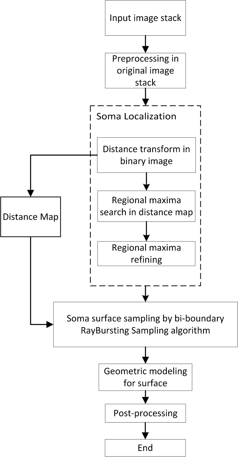
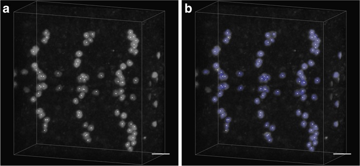
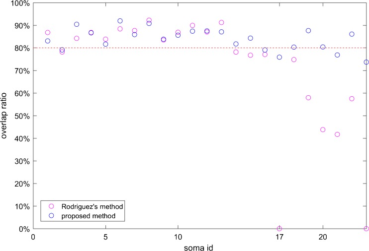
Neuronal soma segmentation is essential for morphology quantification analysis. Rapid advances in light microscope imaging techniques have generated such massive amounts of data that time-consuming manual methods cannot meet requirements for high throughput. However, touching soma segmentation is still a challenge for automatic segmentation methods. In this paper, we propose a soma segmentation method that combines the Rayburst sampling algorithm and ellipsoid fitting. The improved Rayburst sampling algorithm is used to detect the soma surface; the ellipsoid fitting method then refines jagged sampled soma surface to generate smooth ellipsoidal shapes for efficient analysis. In experiments, we validated the proposed method by applying it to datasets from the fluorescence micro-optical sectioning tomography (fMOST) system. The results indicate that the proposed method is comparable to the manual segmented gold standard with accurate soma segmentation at a relatively high speed. The proposed method can be extended to large-scale image stacks in the future.
神经元胞体分割对于形态计量分析至关重要。快速发展的光学显微镜成像技术产生了如此大量的数据,以至于耗时的手动方法无法满足高通量的要求。然而,对于自动分割方法来说,触及胞体分割仍然是一个挑战。在本文中,我们提出了一种结合 Rayburst 采样算法和椭球拟合的胞体分割方法。改进的 Rayburst 采样算法用于检测胞体表面;然后,椭球拟合方法细化锯齿状采样的胞体表面,生成光滑的椭球形状,以便进行有效的分析。在实验中,我们通过将该方法应用于荧光微光学切片层析成像(fMOST)系统的数据集来验证该方法。结果表明,该方法与手动分割的金标准具有可比性,能够以相对较高的速度实现准确的胞体分割。该方法将来可以扩展到大规模的图像堆栈。