• 文献检索
  • 文档翻译
  • 深度研究
  • 学术资讯
  • Suppr Zotero 插件Zotero 插件
  • 邀请有礼
  • 套餐&价格
  • 历史记录
应用&插件
Suppr Zotero 插件Zotero 插件浏览器插件Mac 客户端Windows 客户端微信小程序
定价
高级版会员购买积分包购买API积分包
服务
文献检索文档翻译深度研究API 文档MCP 服务
关于我们
关于 Suppr公司介绍联系我们用户协议隐私条款
关注我们

Suppr 超能文献

核心技术专利:CN118964589B侵权必究
粤ICP备2023148730 号-1Suppr @ 2026

文献检索

告别复杂PubMed语法,用中文像聊天一样搜索,搜遍4000万医学文献。AI智能推荐,让科研检索更轻松。

立即免费搜索

文件翻译

保留排版,准确专业,支持PDF/Word/PPT等文件格式,支持 12+语言互译。

免费翻译文档

深度研究

AI帮你快速写综述,25分钟生成高质量综述,智能提取关键信息,辅助科研写作。

立即免费体验

一种用于分析人睑板腺分泌物的高光谱受激拉曼散射显微镜图像的机器学习框架。

A machine learning framework to analyze hyperspectral stimulated Raman scattering microscopy images of expressed human meibum.

作者信息

Alfonso-García Alba, Paugh Jerry, Farid Marjan, Garg Sumit, Jester James V, Potma Eric O

机构信息

Department of Biomedical Engineering, University of California, Irvine.

Department of Chemistry, University of California, Irvine.

出版信息

J Raman Spectrosc. 2017 Jun;48(6):803-812. doi: 10.1002/jrs.5118. Epub 2017 Apr 11.

DOI:10.1002/jrs.5118
PMID:28943709
原文链接:https://pmc.ncbi.nlm.nih.gov/articles/PMC5608037/
Abstract

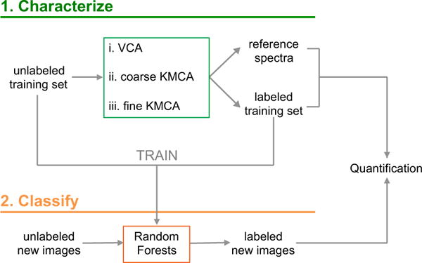
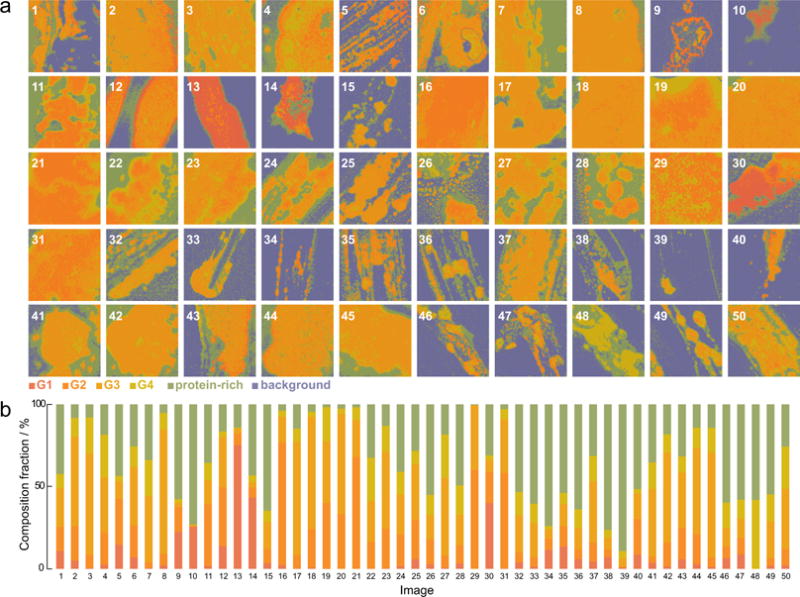
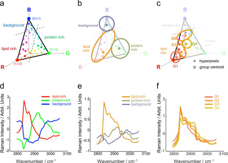
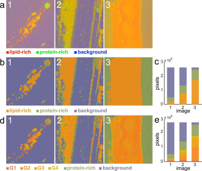
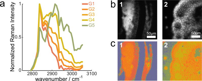
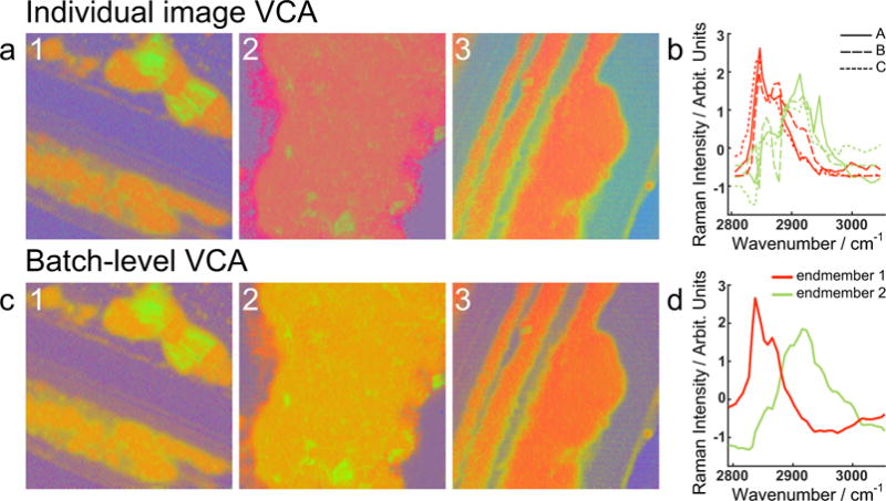
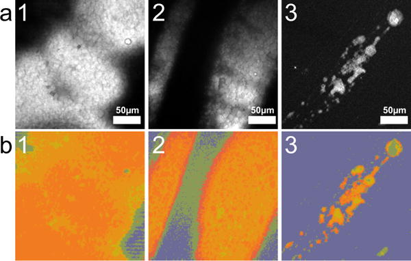
We develop and discuss a methodology for batch-level analysis of hyperspectral stimulated Raman scattering (hsSRS) data sets of human meibum in the CH-stretching vibrational range. The analysis consists of two steps. The first step uses a training set (n=19) to determine chemically meaningful reference spectra that jointly constitute a basis set for the sample. This procedure makes use of batch-level vertex component analysis (VCA), followed by unsupervised k-means clustering to express the data set in terms of spectra that represent lipid and protein mixtures in changing proportions. The second step uses a random forest classifier to rapidly classify hsSRS stacks in terms of the pre-determined basis set. The overall procedure allows a rapid quantitative analysis of large hsSRS data sets, enabling a direct comparison among samples using a single set of reference spectra. We apply this procedure to assess 50 specimens of expressed human meibum, rich in both protein and lipid, and show that the batch-level analysis reveals marked variation among samples that potentially correlate with meibum health quality.

摘要

我们开发并讨论了一种用于批量分析人体睑脂在碳氢键拉伸振动范围内的高光谱受激拉曼散射(hsSRS)数据集的方法。该分析包括两个步骤。第一步使用一个训练集(n = 19)来确定具有化学意义的参考光谱,这些光谱共同构成样本的一个基集。此过程利用批量顶点成分分析(VCA),随后进行无监督k均值聚类,以便根据代表不同比例脂质和蛋白质混合物的光谱来表达数据集。第二步使用随机森林分类器根据预先确定的基集对hsSRS堆栈进行快速分类。整个过程允许对大型hsSRS数据集进行快速定量分析,从而能够使用一组参考光谱对样本进行直接比较。我们应用此程序评估了50个富含蛋白质和脂质的人体睑脂标本,并表明批量分析揭示了样本之间显著的差异,这些差异可能与睑脂健康质量相关。

https://cdn.ncbi.nlm.nih.gov/pmc/blobs/deae/5608037/7afee42653bd/nihms847582f7.jpg
https://cdn.ncbi.nlm.nih.gov/pmc/blobs/deae/5608037/b81206b71955/nihms847582f1.jpg
https://cdn.ncbi.nlm.nih.gov/pmc/blobs/deae/5608037/7c53aad44cbb/nihms847582f2.jpg
https://cdn.ncbi.nlm.nih.gov/pmc/blobs/deae/5608037/046bb6eb7eb2/nihms847582f3.jpg
https://cdn.ncbi.nlm.nih.gov/pmc/blobs/deae/5608037/48d702362e2e/nihms847582f4.jpg
https://cdn.ncbi.nlm.nih.gov/pmc/blobs/deae/5608037/1161dd6e46bf/nihms847582f5.jpg
https://cdn.ncbi.nlm.nih.gov/pmc/blobs/deae/5608037/6520cd407b29/nihms847582f6.jpg
https://cdn.ncbi.nlm.nih.gov/pmc/blobs/deae/5608037/7afee42653bd/nihms847582f7.jpg

相似文献

1
A machine learning framework to analyze hyperspectral stimulated Raman scattering microscopy images of expressed human meibum.一种用于分析人睑板腺分泌物的高光谱受激拉曼散射显微镜图像的机器学习框架。
J Raman Spectrosc. 2017 Jun;48(6):803-812. doi: 10.1002/jrs.5118. Epub 2017 Apr 11.
2
Characterization of expressed human meibum using hyperspectral stimulated Raman scattering microscopy.运用高光谱受激拉曼散射显微镜技术对人眼睑板腺分泌物进行特征描述。
Ocul Surf. 2019 Jan;17(1):151-159. doi: 10.1016/j.jtos.2018.10.003. Epub 2018 Oct 11.
3
Characterization of human meibum lipid using raman spectroscopy.采用拉曼光谱法对人睑板腺脂质进行表征。
Curr Eye Res. 2009 Oct;34(10):824-35. doi: 10.3109/02713680903122029.
4
Broadband hyperspectral stimulated Raman scattering microscopy with a parabolic fiber amplifier source.采用抛物线形光纤放大器源的宽带高光谱受激拉曼散射显微镜
Biomed Opt Express. 2018 Nov 8;9(12):6116-6131. doi: 10.1364/BOE.9.006116. eCollection 2018 Dec 1.
5
Unsupervised hyperspectral stimulated Raman microscopy image enhancement: denoising and segmentation via one-shot deep learning.无监督高光谱受激拉曼显微镜图像增强:通过一次性深度学习进行去噪和分割
Opt Express. 2021 Oct 11;29(21):34205-34219. doi: 10.1364/OE.439662.
6
Efficient quantitative hyperspectral image unmixing method for large-scale Raman micro-spectroscopy data analysis.高效定量高光谱图像解混方法在大规模拉曼显微光谱数据分析中的应用。
Anal Chim Acta. 2019 Mar 7;1050:32-43. doi: 10.1016/j.aca.2018.11.018. Epub 2018 Nov 13.
7
Sparse sampling for fast hyperspectral coherent anti-Stokes Raman scattering imaging.用于快速高光谱相干反斯托克斯拉曼散射成像的稀疏采样
Opt Express. 2014 Feb 24;22(4):4021-8. doi: 10.1364/OE.22.004021.
8
Picosecond spectral coherent anti-Stokes Raman scattering imaging with principal component analysis of meibomian glands.基于主成分分析的皮秒谱相干反斯托克斯拉曼散射成像在睑板腺中的应用。
J Biomed Opt. 2011 Feb;16(2):021104. doi: 10.1117/1.3533716.
9
Development of an image classification pipeline for atherosclerotic plaques assessment using supervised machine learning.应用监督式机器学习开发动脉粥样硬化斑块评估的影像分类流程。
BMC Bioinformatics. 2022 Dec 14;23(1):542. doi: 10.1186/s12859-022-05059-1.
10
Hyperspectral CARS microscopy and quantitative unsupervised analysis of deuterated and non-deuterated fatty acid storage in human cells.高光谱 CARS 显微镜和定量非监督分析人细胞中氘代和非氘代脂肪酸的储存。
J Chem Phys. 2021 Dec 14;155(22):224202. doi: 10.1063/5.0065950.

引用本文的文献

1
Machine learning empowered coherent Raman imaging and analysis for biomedical applications.机器学习助力生物医学应用中的相干拉曼成像与分析。
Commun Eng. 2025 Jan 25;4(1):8. doi: 10.1038/s44172-025-00345-1.
2
Chemical Distribution Uniformity Assessment of "Intra-Tablet" by Hyperspectral Raman Imaging Analysis.采用高光谱拉曼成像分析评估“片内”的化学分布均匀性。
Pharm Res. 2024 Nov;41(11):2247-2258. doi: 10.1007/s11095-024-03778-z. Epub 2024 Nov 6.
3
On machine learning analysis of atomic force microscopy images for image classification, sample surface recognition.

本文引用的文献

1
Comparing Raman and fluorescence lifetime spectroscopy from human atherosclerotic lesions using a bimodal probe.使用双峰探针比较人动脉粥样硬化病变的拉曼光谱和荧光寿命光谱。
J Biophotonics. 2016 Sep;9(9):958-66. doi: 10.1002/jbio.201500341. Epub 2016 Mar 22.
2
Quantitative multi-image analysis for biomedical Raman spectroscopic imaging.用于生物医学拉曼光谱成像的定量多图像分析
J Biophotonics. 2016 May;9(5):542-50. doi: 10.1002/jbio.201500238. Epub 2016 Feb 2.
3
D38-cholesterol as a Raman active probe for imaging intracellular cholesterol storage.
基于原子力显微镜图像的机器学习分析进行图像分类、样本表面识别。
Phys Chem Chem Phys. 2024 Apr 17;26(15):11263-11270. doi: 10.1039/d3cp05673b.
4
Label-Free Imaging of Lipid Droplets in Prostate Cells Using Stimulated Raman Scattering Microscopy and Multivariate Analysis.基于受激拉曼散射显微镜和多元分析的前列腺细胞脂滴无标记成像。
Anal Chem. 2022 Jun 28;94(25):8899-8908. doi: 10.1021/acs.analchem.2c00236. Epub 2022 Jun 14.
5
Quantitative Stimulated Raman Scattering Microscopy: Promises and Pitfalls.定量受激拉曼散射显微镜:前景与挑战。
Annu Rev Anal Chem (Palo Alto Calif). 2022 Jun 13;15(1):269-289. doi: 10.1146/annurev-anchem-061020-015110. Epub 2022 Mar 17.
6
Surface enhanced Raman scattering for the multiplexed detection of pathogenic microorganisms: towards point-of-use applications.基于表面增强拉曼散射的病原体多重检测:迈向现场即时应用。
Analyst. 2021 Oct 11;146(20):6084-6101. doi: 10.1039/d1an00865j.
7
Automatic cell counting from stimulated Raman imaging using deep learning.基于深度学习的受激拉曼成像的自动细胞计数。
PLoS One. 2021 Jul 21;16(7):e0254586. doi: 10.1371/journal.pone.0254586. eCollection 2021.
8
Mammalian cell and tissue imaging using Raman and coherent Raman microscopy.使用拉曼和相干拉曼显微镜进行哺乳动物细胞和组织成像。
Wiley Interdiscip Rev Syst Biol Med. 2020 Nov;12(6):e1501. doi: 10.1002/wsbm.1501. Epub 2020 Jul 19.
9
Eicosapentaenoic acid (EPA) activates PPARγ signaling leading to cell cycle exit, lipid accumulation, and autophagy in human meibomian gland epithelial cells (hMGEC).二十碳五烯酸(EPA)激活过氧化物酶体增殖物激活受体γ(PPARγ)信号通路,导致人睑板腺上皮细胞(hMGEC)退出细胞周期、脂质蓄积和自噬。
Ocul Surf. 2020 Jul;18(3):427-437. doi: 10.1016/j.jtos.2020.04.012. Epub 2020 Apr 30.
10
Machine Learning based Analytical Framework for Automatic Hyperspectral Raman Analysis of Lithium-ion Battery Electrodes.基于机器学习的锂离子电池电极自动高光谱拉曼分析框架
Sci Rep. 2019 Dec 3;9(1):18241. doi: 10.1038/s41598-019-54770-2.
D38-胆固醇作为用于成像细胞内胆固醇储存的拉曼活性探针。
J Biomed Opt. 2016 Jun;21(6):61003. doi: 10.1117/1.JBO.21.6.061003.
4
Spectrometer-free vibrational imaging by retrieving stimulated Raman signal from highly scattered photons.通过从高度散射光子中检索受激拉曼信号实现无光谱仪振动成像。
Sci Adv. 2015 Oct 30;1(9):e1500738. doi: 10.1126/sciadv.1500738. eCollection 2015 Oct.
5
Label-free DNA imaging in vivo with stimulated Raman scattering microscopy.利用受激拉曼散射显微镜进行体内无标记DNA成像。
Proc Natl Acad Sci U S A. 2015 Sep 15;112(37):11624-9. doi: 10.1073/pnas.1515121112. Epub 2015 Aug 31.
6
Microsecond Scale Vibrational Spectroscopic Imaging by Multiplex Stimulated Raman Scattering Microscopy.基于多重受激拉曼散射显微镜的微秒级振动光谱成像
Light Sci Appl. 2015;4. doi: 10.1038/lsa.2015.38.
7
Multivariate Curve Resolution Applied to Hyperspectral Imaging Analysis of Chocolate Samples.多元曲线分辨法应用于巧克力样品的高光谱成像分析
Appl Spectrosc. 2015 Aug;69(8):993-1003. doi: 10.1366/14-07819. Epub 2015 Jul 1.
8
Raman imaging providing insights into chemical composition of lipid droplets of different size and origin: in hepatocytes and endothelium.拉曼成像提供了对不同大小和来源的脂滴的化学成分的深入了解:在肝细胞和内皮细胞中。
Anal Chem. 2014 Jul 1;86(13):6666-74. doi: 10.1021/ac501395g. Epub 2014 Jun 17.
9
Automated identification of subcellular organelles by coherent anti-stokes Raman scattering.通过相干反斯托克斯拉曼散射自动识别亚细胞器
Biophys J. 2014 May 6;106(9):1910-20. doi: 10.1016/j.bpj.2014.03.025.
10
Reliable cell segmentation based on spectral phasor analysis of hyperspectral stimulated Raman scattering imaging data.基于高光谱受激拉曼散射成像数据的光谱相衬分析的可靠细胞分割。
Anal Chem. 2014 May 6;86(9):4115-9. doi: 10.1021/ac500014b. Epub 2014 Apr 8.