• 文献检索
  • 文档翻译
  • 深度研究
  • 学术资讯
  • Suppr Zotero 插件Zotero 插件
  • 邀请有礼
  • 套餐&价格
  • 历史记录
应用&插件
Suppr Zotero 插件Zotero 插件浏览器插件Mac 客户端Windows 客户端微信小程序
定价
高级版会员购买积分包购买API积分包
服务
文献检索文档翻译深度研究API 文档MCP 服务
关于我们
关于 Suppr公司介绍联系我们用户协议隐私条款
关注我们

Suppr 超能文献

核心技术专利:CN118964589B侵权必究
粤ICP备2023148730 号-1Suppr @ 2026

文献检索

告别复杂PubMed语法,用中文像聊天一样搜索,搜遍4000万医学文献。AI智能推荐,让科研检索更轻松。

立即免费搜索

文件翻译

保留排版,准确专业,支持PDF/Word/PPT等文件格式,支持 12+语言互译。

免费翻译文档

深度研究

AI帮你快速写综述,25分钟生成高质量综述,智能提取关键信息,辅助科研写作。

立即免费体验

无皮层入侵的内侧前额叶皮层和海马体的双光子钙成像。

Two-photon calcium imaging of the medial prefrontal cortex and hippocampus without cortical invasion.

机构信息

Department of Physiology, Graduate School of Medicine, The University of Tokyo, Tokyo, Japan.

Division of Brain Circuits, National Institute for Basic Biology, Okazaki, Japan.

出版信息

Elife. 2017 Sep 25;6:e26839. doi: 10.7554/eLife.26839.

DOI:10.7554/eLife.26839
PMID:28945191
原文链接:https://pmc.ncbi.nlm.nih.gov/articles/PMC5643091/
Abstract

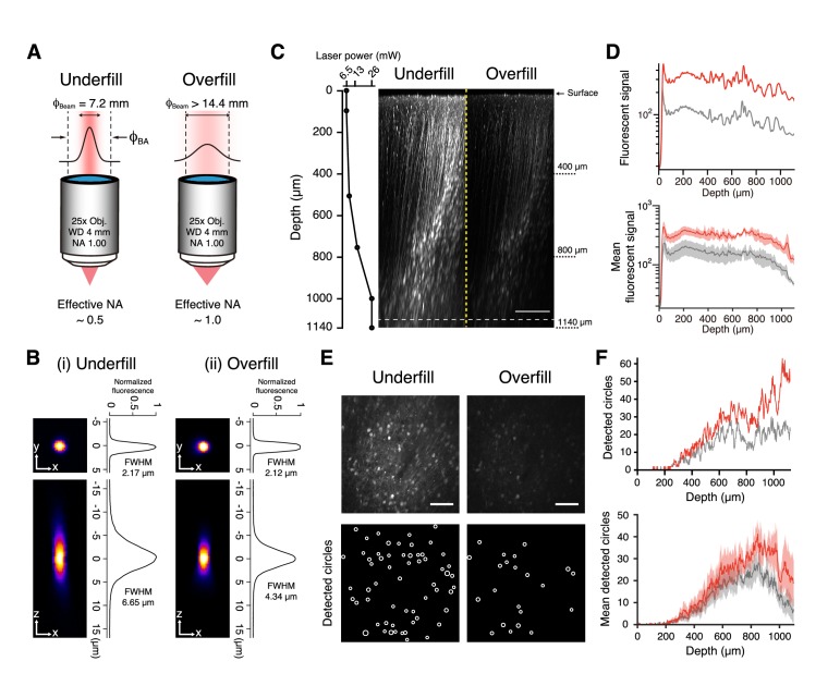
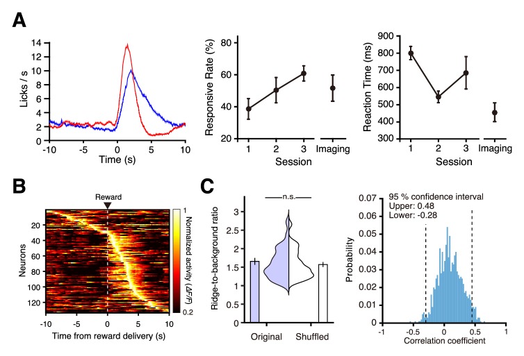
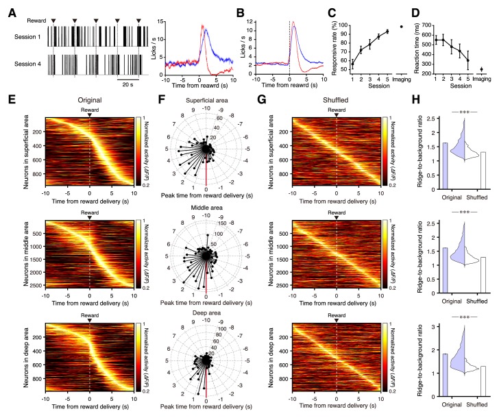
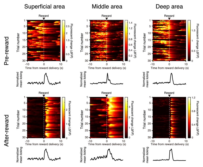
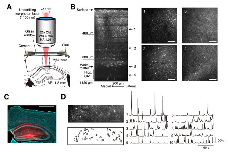
In vivo two-photon calcium imaging currently allows us to observe the activity of multiple neurons up to ~900 µm below the cortical surface without cortical invasion. However, many important brain areas are located deeper than this. Here, we used an 1100 nm laser that underfilled the back aperture of the objective together with red genetically encoded calcium indicators to establish two-photon calcium imaging of the intact mouse brain and detect neural activity up to 1200 μm from the cortical surface. This imaging was obtained from the medial prefrontal cortex (the prelimbic area) and the hippocampal CA1 region. We found that neural activity before water delivery repeated at a constant interval was higher in the prelimbic area than in layer 2/3 of the secondary motor area. Reducing the invasiveness of imaging is an important strategy to reveal the intact brain processes active in cognition and memory.

摘要

在体双光子钙成像目前允许我们在不侵犯皮层的情况下观察皮层下约 900 µm 深度处的多个神经元的活动。然而,许多重要的脑区位于更深的位置。在这里,我们使用了一种 1100nm 的激光,该激光使物镜的后孔径未完全充满,同时使用红色遗传编码钙指示剂,建立了完整小鼠大脑的双光子钙成像,并从皮层表面检测到了 1200μm 深度的神经活动。这种成像来自于内侧前额叶皮层(前额叶区域)和海马 CA1 区域。我们发现,在水输送之前,重复出现的神经活动在前额叶区域比在次级运动区的第 2/3 层更高。降低成像的侵入性是揭示在认知和记忆中活跃的完整大脑过程的重要策略。

https://cdn.ncbi.nlm.nih.gov/pmc/blobs/f76f/5643091/eb031b4ad2dc/elife-26839-fig6.jpg
https://cdn.ncbi.nlm.nih.gov/pmc/blobs/f76f/5643091/57b329f4dbb3/elife-26839-fig1.jpg
https://cdn.ncbi.nlm.nih.gov/pmc/blobs/f76f/5643091/1500d8ab6ff5/elife-26839-fig2.jpg
https://cdn.ncbi.nlm.nih.gov/pmc/blobs/f76f/5643091/76c670d99010/elife-26839-fig3.jpg
https://cdn.ncbi.nlm.nih.gov/pmc/blobs/f76f/5643091/d6129b3d87c0/elife-26839-fig3-figsupp1.jpg
https://cdn.ncbi.nlm.nih.gov/pmc/blobs/f76f/5643091/8befb04ea3c3/elife-26839-fig3-figsupp2.jpg
https://cdn.ncbi.nlm.nih.gov/pmc/blobs/f76f/5643091/0497df06980c/elife-26839-fig3-figsupp3.jpg
https://cdn.ncbi.nlm.nih.gov/pmc/blobs/f76f/5643091/3f8b3fd09edc/elife-26839-fig4.jpg
https://cdn.ncbi.nlm.nih.gov/pmc/blobs/f76f/5643091/43cc2e98441e/elife-26839-fig4-figsupp1.jpg
https://cdn.ncbi.nlm.nih.gov/pmc/blobs/f76f/5643091/e77715f2229e/elife-26839-fig4-figsupp2.jpg
https://cdn.ncbi.nlm.nih.gov/pmc/blobs/f76f/5643091/7e5d7a06cad4/elife-26839-fig4-figsupp3.jpg
https://cdn.ncbi.nlm.nih.gov/pmc/blobs/f76f/5643091/2a193f1c27b5/elife-26839-fig4-figsupp4.jpg
https://cdn.ncbi.nlm.nih.gov/pmc/blobs/f76f/5643091/654a3d4177a4/elife-26839-fig5.jpg
https://cdn.ncbi.nlm.nih.gov/pmc/blobs/f76f/5643091/827e70f4366f/elife-26839-fig5-figsupp1.jpg
https://cdn.ncbi.nlm.nih.gov/pmc/blobs/f76f/5643091/eb031b4ad2dc/elife-26839-fig6.jpg

相似文献

1
Two-photon calcium imaging of the medial prefrontal cortex and hippocampus without cortical invasion.无皮层入侵的内侧前额叶皮层和海马体的双光子钙成像。
Elife. 2017 Sep 25;6:e26839. doi: 10.7554/eLife.26839.
2
A large field of view two-photon mesoscope with subcellular resolution for in vivo imaging.一种用于体内成像的具有亚细胞分辨率的大视野双光子显微镜。
Elife. 2016 Jun 14;5:e14472. doi: 10.7554/eLife.14472.
3
Topographically specific hippocampal projections target functionally distinct prefrontal areas in the rhesus monkey.在恒河猴中,海马的拓扑特异性投射靶向功能不同的前额叶区域。
Hippocampus. 1995;5(6):511-33. doi: 10.1002/hipo.450050604.
4
Hippocampo-prefrontal cortex pathway: anatomical and electrophysiological characteristics.海马-前额叶皮质通路:解剖学和电生理特征
Hippocampus. 2000;10(4):411-9. doi: 10.1002/1098-1063(2000)10:4<411::AID-HIPO7>3.0.CO;2-A.
5
In Vivo Two-Photon Calcium Imaging of Hippocampal Neurons in Alzheimer Mouse Models.阿尔茨海默病小鼠模型中海马神经元的体内双光子钙成像
Methods Mol Biol. 2018;1750:341-351. doi: 10.1007/978-1-4939-7704-8_23.
6
High-yield in vitro recordings from neurons functionally characterized in vivo.从体内功能特征化的神经元中获得高产的体外记录。
Nat Protoc. 2018 Jun;13(6):1275-1293. doi: 10.1038/nprot.2018.026. Epub 2018 May 10.
7
Chronic calcium imaging of neurons in the mouse visual cortex using a troponin C-based indicator.使用基于肌钙蛋白C的指示剂对小鼠视觉皮层神经元进行慢性钙成像。
Cold Spring Harb Protoc. 2014 May 1;2014(5):pdb.prot081737. doi: 10.1101/pdb.prot081737.
8
Corticotropin-releasing factor infusion into nucleus incertus suppresses medial prefrontal cortical activity and hippocampo-medial prefrontal cortical long-term potentiation.促肾上腺皮质素释放因子输注到中脑导水管周围灰质抑制前额皮质内侧活动和海马-前额皮质内侧长时程增强。
Eur J Neurosci. 2013 Aug;38(4):2516-25. doi: 10.1111/ejn.12242. Epub 2013 May 14.
9
Intravital FRET: Probing Cellular and Tissue Function in Vivo.活体荧光共振能量转移:探究体内细胞和组织功能
Int J Mol Sci. 2015 May 21;16(5):11713-27. doi: 10.3390/ijms160511713.
10
Disconnection of the hippocampal-prefrontal cortical circuits impairs spatial working memory performance in rats.海马体-前额叶皮质回路的切断会损害大鼠的空间工作记忆表现。
Behav Brain Res. 2006 Dec 15;175(2):329-36. doi: 10.1016/j.bbr.2006.09.002. Epub 2006 Oct 10.

引用本文的文献

1
Advances in two-photon imaging for monitoring neural activity in behaving mice.用于监测行为小鼠神经活动的双光子成像技术进展。
Front Neurosci. 2025 Aug 11;19:1597151. doi: 10.3389/fnins.2025.1597151. eCollection 2025.
2
Compressive Fourier-Domain Intensity Coupling (C-FOCUS) enables near-millimeter deep imaging in the intact mouse brain in vivo.压缩傅里叶域强度耦合(C-FOCUS)能够在完整的活体小鼠大脑中实现近毫米深度成像。
Res Sq. 2025 Jul 28:rs.3.rs-7104566. doi: 10.21203/rs.3.rs-7104566/v1.
3
Scattering correction through Fourier-domain intensity coupling in two-photon microscopy (2P-FOCUS).

本文引用的文献

1
Prefrontal cortex output circuits guide reward seeking through divergent cue encoding.前额叶皮质输出回路通过不同的线索编码引导奖赏寻求行为。
Nature. 2017 Mar 2;543(7643):103-107. doi: 10.1038/nature21376. Epub 2017 Feb 22.
2
In vivo three-photon imaging of activity of GCaMP6-labeled neurons deep in intact mouse brain.在完整小鼠大脑深处用 GCaMP6 标记的神经元的体内三光子成像。
Nat Methods. 2017 Apr;14(4):388-390. doi: 10.1038/nmeth.4183. Epub 2017 Feb 20.
3
Long-Term Optical Access to an Estimated One Million Neurons in the Live Mouse Cortex.
通过双光子显微镜(2P-FOCUS)中的傅里叶域强度耦合进行散射校正。
Photonics Res. 2025 Apr;13(4):845-864. doi: 10.1364/prj.544387. Epub 2025 Mar 11.
4
Three-photon in vivo imaging of neurons and glia in the medial prefrontal cortex with sub-cellular resolution.内侧前额叶皮质中神经元和神经胶质细胞的三光子亚细胞分辨率体内成像。
Commun Biol. 2025 May 23;8(1):795. doi: 10.1038/s42003-025-08079-8.
5
Whether or not to act is determined by distinct signals from motor thalamus and orbitofrontal cortex to secondary motor cortex.是否采取行动由来自运动丘脑和眶额皮质至次级运动皮质的不同信号决定。
Nat Commun. 2025 Apr 4;16(1):3106. doi: 10.1038/s41467-025-58272-w.
6
Standardized measurements for monitoring and comparing multiphoton microscope systems.用于监测和比较多光子显微镜系统的标准化测量方法。
Nat Protoc. 2025 Mar 17. doi: 10.1038/s41596-024-01120-w.
7
Scattering Correction through Fourier-Domain Intensity Coupling in Two-Photon Microscopy (2P-FOCUS).通过双光子显微镜(2P-FOCUS)中的傅里叶域强度耦合进行散射校正
ArXiv. 2025 Feb 10:arXiv:2401.15192v2.
8
Out-of-focus signal rejection for pO measurements using two-photon phosphorescence lifetime microscopy.使用双光子磷光寿命显微镜进行pO测量时的离焦信号抑制
Biomed Opt Express. 2024 Dec 12;16(1):159-176. doi: 10.1364/BOE.532084. eCollection 2025 Jan 1.
9
Calcium Indicators with Fluorescence Lifetime-Based Signal Readout: A Structure-Function Study.基于荧光寿命信号读出的钙指示剂:结构-功能研究
Int J Mol Sci. 2024 Nov 21;25(23):12493. doi: 10.3390/ijms252312493.
10
Multiphoton fluorescence microscopy for in vivo imaging.多光子荧光显微镜用于活体成像。
Cell. 2024 Aug 22;187(17):4458-4487. doi: 10.1016/j.cell.2024.07.036.
长期光学接入活体小鼠皮层中估计达一百万个神经元。
Cell Rep. 2016 Dec 20;17(12):3385-3394. doi: 10.1016/j.celrep.2016.12.004.
4
Survival of corticostriatal neurons by Rho/Rho-kinase signaling pathway.通过Rho/ Rho激酶信号通路实现皮质纹状体神经元的存活。
Neurosci Lett. 2016 Sep 6;630:45-52. doi: 10.1016/j.neulet.2016.07.020. Epub 2016 Jul 15.
5
Functional Imaging of Dentate Granule Cells in the Adult Mouse Hippocampus.成年小鼠海马齿状颗粒细胞的功能成像
J Neurosci. 2016 Jul 13;36(28):7407-14. doi: 10.1523/JNEUROSCI.3065-15.2016.
6
Wide field-of-view, multi-region, two-photon imaging of neuronal activity in the mammalian brain.哺乳动物大脑神经元活动的宽视野、多区域双光子成像。
Nat Biotechnol. 2016 Aug;34(8):857-62. doi: 10.1038/nbt.3594. Epub 2016 Jun 27.
7
A large field of view two-photon mesoscope with subcellular resolution for in vivo imaging.一种用于体内成像的具有亚细胞分辨率的大视野双光子显微镜。
Elife. 2016 Jun 14;5:e14472. doi: 10.7554/eLife.14472.
8
Brain heating induced by near-infrared lasers during multiphoton microscopy.多光子显微镜检查期间近红外激光引起的脑部发热
J Neurophysiol. 2016 Sep 1;116(3):1012-23. doi: 10.1152/jn.00275.2016. Epub 2016 Jun 8.
9
Over the river, through the woods: cognitive maps in the hippocampus and orbitofrontal cortex.越过河流,穿过树林:海马体与眶额皮质中的认知地图
Nat Rev Neurosci. 2016 Aug;17(8):513-23. doi: 10.1038/nrn.2016.56. Epub 2016 Jun 3.
10
Sensitive red protein calcium indicators for imaging neural activity.用于神经活动成像的灵敏红色蛋白质钙指示剂。
Elife. 2016 Mar 24;5:e12727. doi: 10.7554/eLife.12727.