Wang Wen-Ke, Lu Qing-Hua, Wang Xin, Wang Ben, Wang Juan, Gong Hui-Ping, Wang Lin, Li Hao, Du Yi-Meng
Department of Cardiology, The Second Hospital of Shandong University, Jinan, Shandong 250033, P.R. China.
Department of General Surgery, Qilu Hospital of Shandong University, Jinan, Shandong 250012, P.R. China.
Exp Ther Med. 2017 Sep;14(3):2497-2504. doi: 10.3892/etm.2017.4824. Epub 2017 Jul 19.
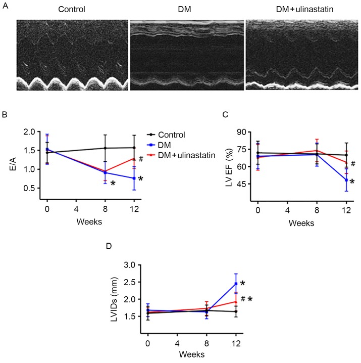
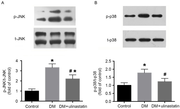
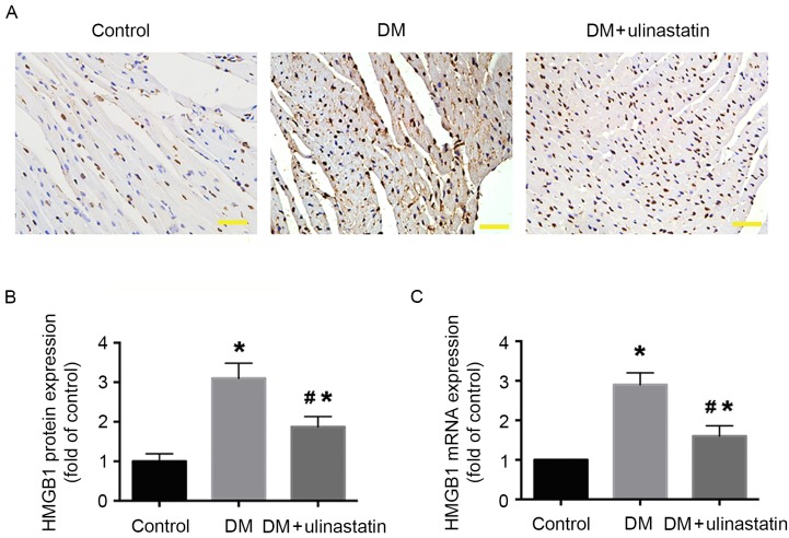
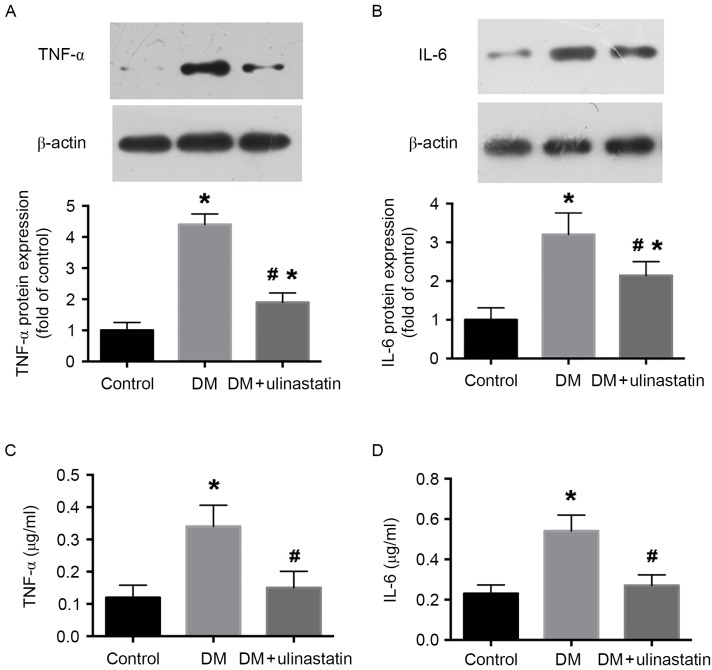
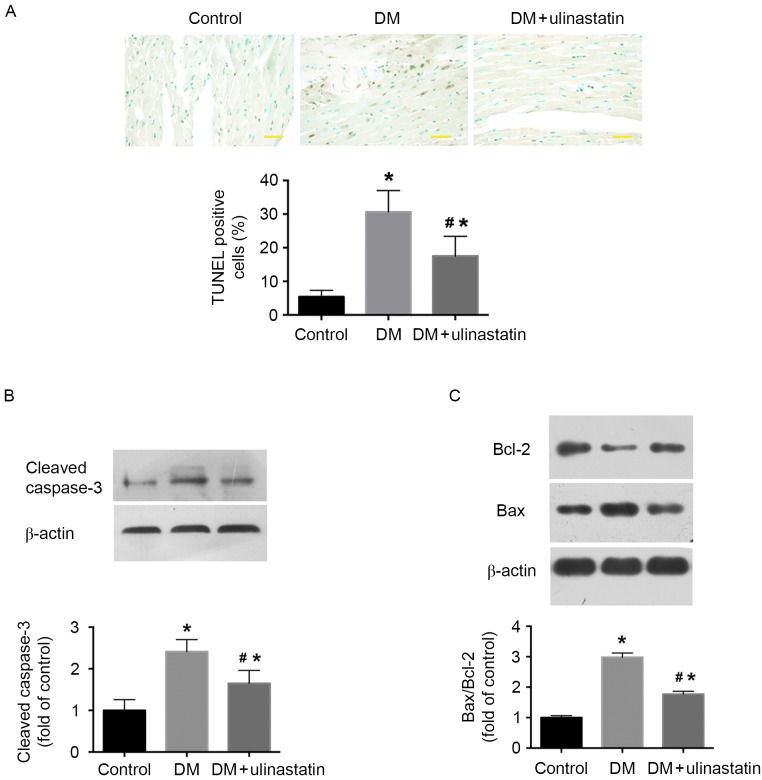
Ulinastatin exhibits anti-inflammatory activity and protects the heart from ischemia/reperfusion injury. However, whether ulinastatin has a protective effect in diabetic cardiomyopathy is yet to be elucidated. The aim of the present study was to investigate the protective effects of ulinastatin against diabetic cardiomyopathy and its underlying mechanisms. A C57/BL6J mice model of diabetic cardiomyopathy was used and mice were randomly assigned to three groups: Control group, diabetes mellitus (DM) group and DM + ulinastatin treatment group. Cardiac function was assessed using echocardiography and the level of inflammatory cytokine high mobility group box 1 (HMGB1) expression was measured using histopathological examination and reverse transcription-quantitative polymerase chain reaction. The levels of tumor necrosis factor (TNF)-α and interleukin (IL)-6 were measured using western blotting and ELISA. The apoptosis rate in the myocardium was assessed by TUNEL assay. Caspase-3 activation, expression of B-cell lymphoma 2 (Bcl-2) and Bcl-2 associated × (Bax) were measured using western blotting, as was the activity of the mitogen activated protein kinase (MAPK) signaling pathway. The results indicated that ulinastatin significantly improved cardiac function in mice with DM. Ulinastatin treatment significantly downregulated HMGB1, TNF-α and IL-6 expression (P<0.05) and significantly reduced the percentage of apoptotic cardiomyocytes (P<0.05) via reduction of caspase-3 activation and the ratio of Bax/Bcl-2 in diabetic hearts (P<0.05). In addition, ulinastatin attenuated the activation of the MAPK signaling pathway. In conclusion, ulinastatin had a protective effect against DM-induced cardiac dysfunction in a mouse model. This protective effect may be associated with the anti-inflammatory and anti-apoptotic abilities of ulinastatin via the MAPK signaling pathway.
乌司他丁具有抗炎活性,并能保护心脏免受缺血/再灌注损伤。然而,乌司他丁在糖尿病性心肌病中是否具有保护作用尚待阐明。本研究的目的是探讨乌司他丁对糖尿病性心肌病的保护作用及其潜在机制。采用C57/BL6J小鼠糖尿病性心肌病模型,将小鼠随机分为三组:对照组、糖尿病(DM)组和DM+乌司他丁治疗组。采用超声心动图评估心脏功能,采用组织病理学检查和逆转录-定量聚合酶链反应检测炎症细胞因子高迁移率族蛋白B1(HMGB1)的表达水平。采用蛋白质印迹法和酶联免疫吸附测定法检测肿瘤坏死因子(TNF)-α和白细胞介素(IL)-6的水平。通过TUNEL法评估心肌细胞凋亡率。采用蛋白质印迹法检测半胱天冬酶-3的激活、B细胞淋巴瘤2(Bcl-2)和Bcl-2相关X蛋白(Bax)的表达,以及丝裂原活化蛋白激酶(MAPK)信号通路的活性。结果表明,乌司他丁显著改善了糖尿病小鼠的心脏功能。乌司他丁治疗显著下调了HMGB1、TNF-α和IL-6的表达(P<0.05),并通过降低糖尿病心脏中半胱天冬酶-3的激活以及Bax/Bcl-2的比值,显著降低了凋亡心肌细胞的百分比(P<0.05)。此外,乌司他丁减弱了MAPK信号通路的激活。总之,在小鼠模型中,乌司他丁对糖尿病诱导的心脏功能障碍具有保护作用。这种保护作用可能与乌司他丁通过MAPK信号通路的抗炎和抗凋亡能力有关。