Schecklmann Martin, Mann Alexander, Langguth Berthold, Ehlis Ann-Christine, Fallgatter Andreas J, Haeussinger Florian B
Department of Psychiatry and Psychotherapy, University of RegensburgRegensburg, Germany.
Department of Psychiatry and Psychotherapy, Psychophysiology and Optical Imaging, University Hospital of TübingenTübingen, Germany.
Front Hum Neurosci. 2017 Sep 15;11:456. doi: 10.3389/fnhum.2017.00456. eCollection 2017.
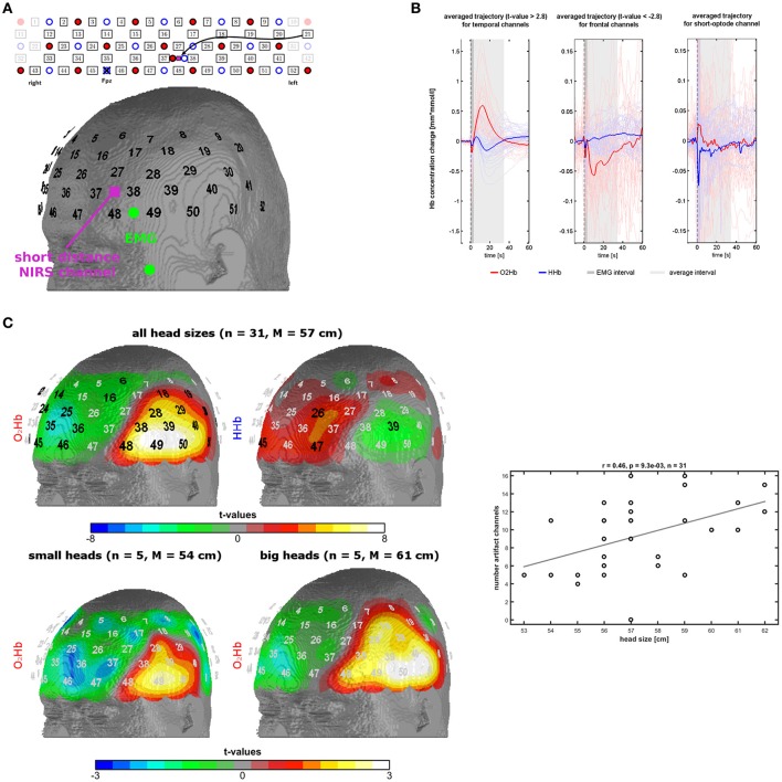
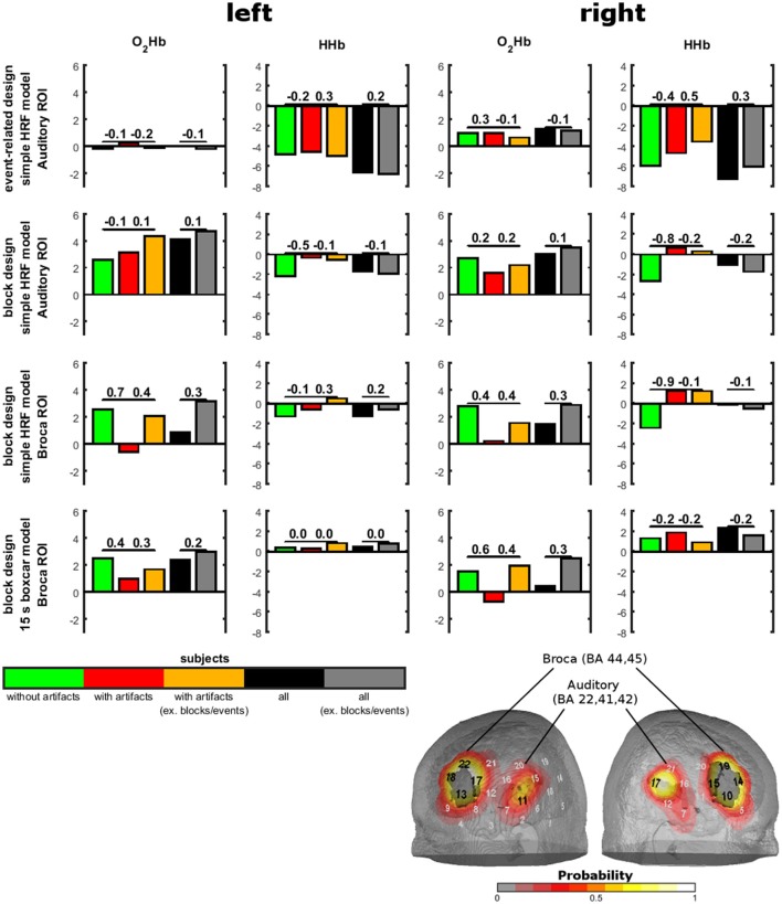
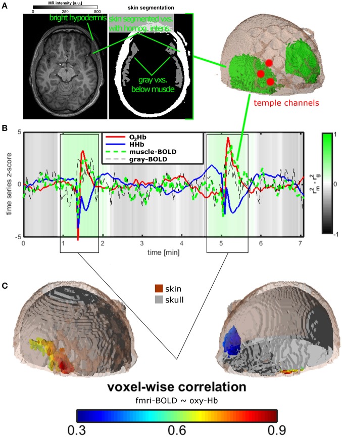
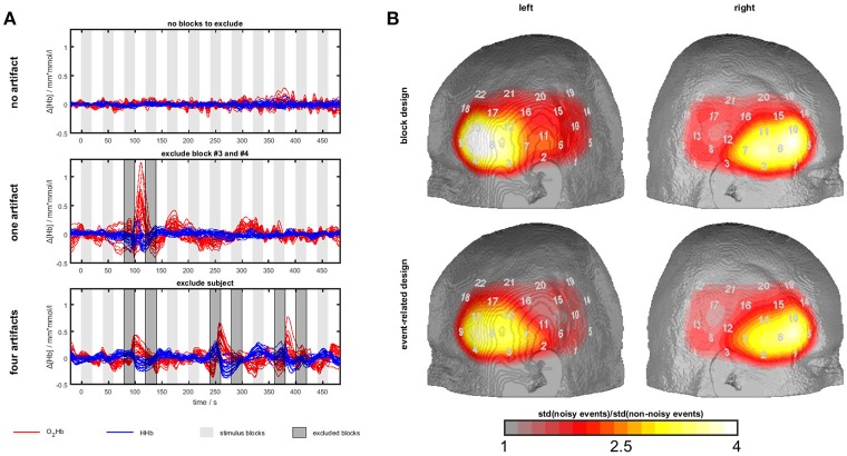
Extracranial signals are the main source of noise in functional near-infrared spectroscopy (fNIRS) as light is penetrating the cortex but also skin and muscles of the head. Here we performed three experiments to investigate the contamination of fNIRS measurements by temporal muscle activity. For experiment 1, we provoked temporal muscle activity by instructing 31 healthy subjects to clench their teeth three times. We measured fNIRS signals over left temporal and frontal channels with an interoptode distance of 3 cm, in one short optode distance (SOD) channel (1 cm) and electromyography (EMG) over the edge of the temporal muscle. In experiment 2, we screened resting state fNIRS-fMRI (functional magnetic resonance imaging) data of one healthy subject for temporal muscle artifacts. In experiment 3, we screened a dataset of sound-evoked activity ( = 33) using bi-temporal probe-sets and systematically contrasted subjects presenting vs. not presenting artifacts and blocks/events contaminated or not contaminated with artifacts. In experiment 1, we could demonstrate a hemodynamic-response-like increase in oxygenated (OHb) and decrease in deoxygenated (HHb) hemoglobin with a large amplitude and large spatial extent highly exceeding normal cortical activity. Correlations between EMG, SOD, and fNIRS artifact activity showed only limited evidence for associations on a group level with rather clear associations in a sub-group of subjects. The fNIRS-fMRI experiment showed that during the temporal muscle artifact, fNIRS is completely saturated by muscle oxygenation. Experiment 3 showed hints for contamination of sound-evoked oxygenation by the temporal muscle artifact. This was of low relevance in analyzing the whole sample. Temporal muscle activity e.g., by clenching the teeth induces a large hemodynamic-like artifact in fNIRS measurements which should be avoided by specific subject instructions. Data should be screened for this artifact might be corrected by exclusion of contaminated blocks/events. The usefulness of established artifact correction methods should be evaluated in future studies. Temporal muscle activity, e.g., by clenching the teeth is one major source of noise in fNIRS measurements.
颅外信号是功能近红外光谱(fNIRS)中噪声的主要来源,因为光线穿透皮层的同时也穿透头部的皮肤和肌肉。在此,我们进行了三项实验,以研究颞肌活动对fNIRS测量的干扰。在实验1中,我们指示31名健康受试者咬紧牙关三次,以引发颞肌活动。我们在左颞叶和额叶通道上测量fNIRS信号,光极间距为3 cm,在一个短光极间距(SOD)通道(1 cm)以及颞肌边缘的肌电图(EMG)。在实验2中,我们筛查了一名健康受试者的静息态fNIRS - fMRI(功能磁共振成像)数据中的颞肌伪影。在实验3中,我们使用双侧颞部探头组筛查了一个声音诱发活动的数据集(n = 33),并系统地对比了出现与未出现伪影以及受伪影污染或未受污染的组块/事件的受试者。在实验1中,我们可以证明,氧合血红蛋白(OHb)出现类似血流动力学反应的增加,而脱氧血红蛋白(HHb)减少,其幅度大且空间范围广,远超正常皮层活动。EMG、SOD和fNIRS伪影活动之间的相关性在组水平上仅显示出有限的关联证据,而在一个亚组受试者中关联较为明显。fNIRS - fMRI实验表明,在颞肌伪影期间,fNIRS完全被肌肉氧合饱和。实验3显示出颞肌伪影对声音诱发氧合有干扰的迹象。这在分析整个样本时相关性较低。例如,通过咬紧牙关产生的颞肌活动会在fNIRS测量中诱发类似血流动力学的大伪影,应通过特定的受试者指导来避免。对于该伪影,数据应进行筛查,可能通过排除受污染的组块/事件来校正。已建立的伪影校正方法的有效性应在未来研究中进行评估。例如,通过咬紧牙关产生的颞肌活动是fNIRS测量中一个主要的噪声来源。