Department of Veterinary Physiology, College of Veterinary Medicine, Research Institute for Veterinary Science, and BK21 PLUS program for Creative Veterinary Research Center, Seoul National University, Seoul, 08826, South Korea.
Department of Agricultural Biotechnology, Animal Biotechnology Major, and Research Institute for Agriculture and Life science, Seoul National University, Seoul, 08826, South Korea.
Sci Rep. 2017 Oct 3;7(1):12582. doi: 10.1038/s41598-017-12692-x.
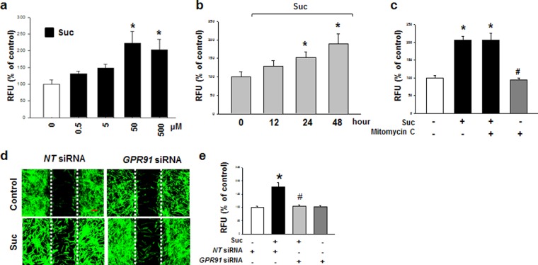
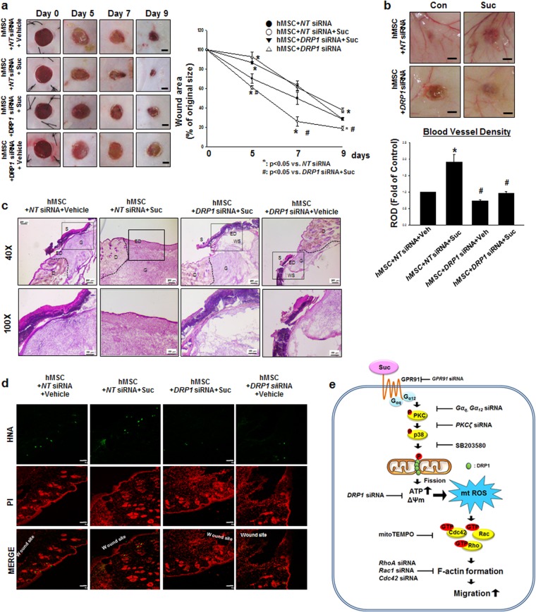
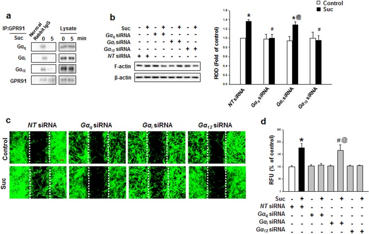
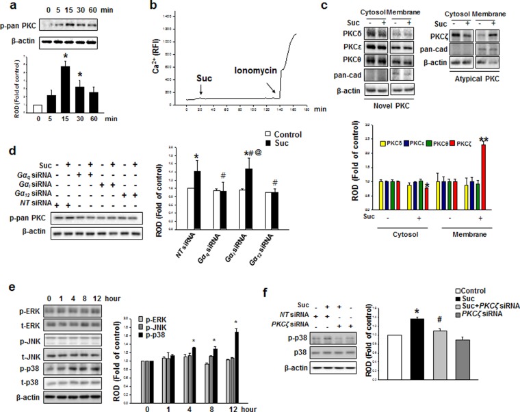
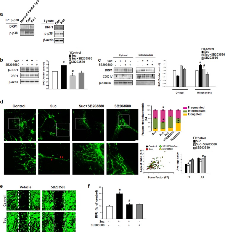
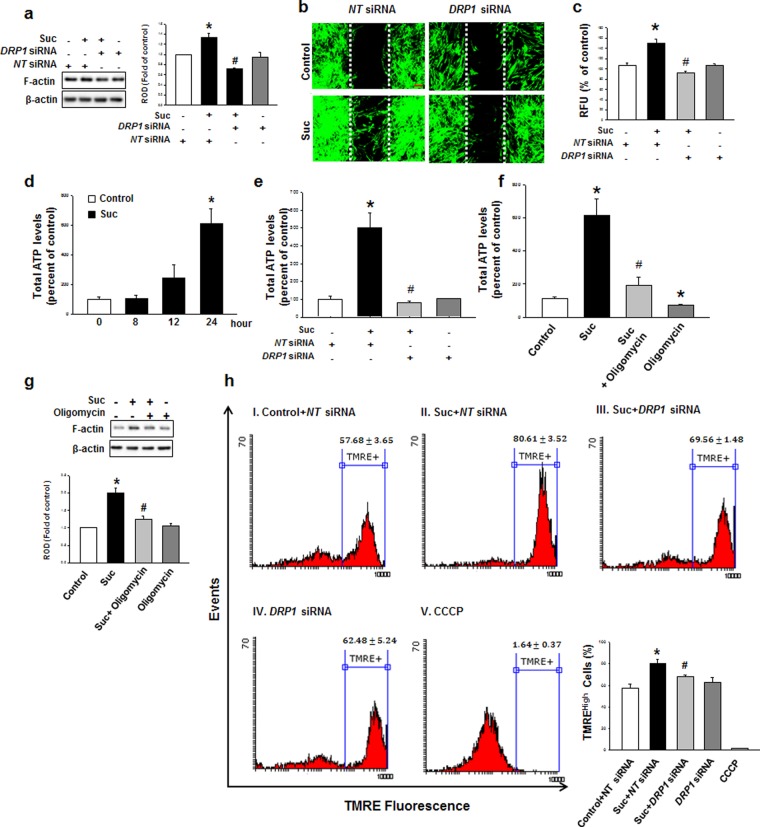
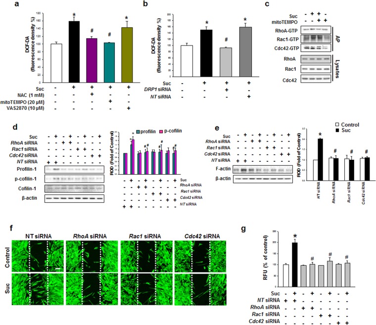
The role of metabolites produced from stem cell metabolism has been emerged as signaling molecules to regulate stem cell behaviors such as migration. The mitochondrial morphology is closely associated with the metabolic balance and stem cell function. However, the physiological role of succinate on human mesenchymal stem cell (hMSC) migration by regulating the mitochondrial morphology remains unclear. Here, we investigate the effect of succinate on hMSC migration via regulation of mitochondrial dynamics and its related signaling pathway. Succinate (50 μM) significantly accelerates hMSC migration. Succinate increases phosphorylation of pan-PKC, especially the atypical PKCζ level which was blocked by the knockdown of Gα and Gα Activated PKCζ subsequently phosphorylates p38 MAPK. Cytosolic DRP1 is phosphorylated by p38 MAPK and results in DRP1 translocation to the mitochondria outer membrane, eventually inducing mitochondrial fragmentation. Mitochondrial fission-induced mitochondrial function elevates mitochondrial ROS (mtROS) levels and activates Rho GTPases, which then induces F-actin formation. Furthermore, in a skin excisional wound model, we found the effects of succinate-pretreated hMSC enhanced wound closure, vascularization and re-epithelialization and confirmed that DRP1 has a vital role in injured tissue regeneration. Overall, succinate promotes DRP1-mediated mitochondrial fission via GPR91, consequently stimulating the hMSC migration through mtROS-induced F-actin formation.
干细胞代谢产生的代谢物作为信号分子的作用已经显现出来,可以调节干细胞的行为,如迁移。线粒体形态与代谢平衡和干细胞功能密切相关。然而,琥珀酸通过调节线粒体形态对人间充质干细胞(hMSC)迁移的生理作用尚不清楚。在这里,我们通过调节线粒体动力学及其相关信号通路来研究琥珀酸对 hMSC 迁移的影响。琥珀酸(50μM)显著加速 hMSC 迁移。琥珀酸增加了全 PKC 的磷酸化,特别是非典型 PKCζ 水平,这一水平被 Gα 和 Gα 激活的 PKCζ 的敲低所阻断,随后磷酸化 p38 MAPK。细胞质中的 DRP1 被 p38 MAPK 磷酸化,导致 DRP1 转位到线粒体外膜,最终诱导线粒体碎片化。线粒体分裂诱导的线粒体功能提高了线粒体 ROS(mtROS)水平并激活了 Rho GTPases,从而诱导 F-肌动蛋白的形成。此外,在皮肤切除性伤口模型中,我们发现经琥珀酸预处理的 hMSC 的作用增强了伤口闭合、血管生成和再上皮化,并证实 DRP1 在受损组织再生中起着至关重要的作用。总的来说,琥珀酸通过 GPR91 促进 DRP1 介导的线粒体分裂,从而通过 mtROS 诱导的 F-肌动蛋白形成刺激 hMSC 迁移。