Lindberg Olof, Mårtensson Gustav, Stomrud Erik, Palmqvist Sebastian, Wahlund Lars-Olof, Westman Eric, Hansson Oskar
Clinical Memory Research Unit, Department of Clinical Sciences, Lund UniversityLund, Sweden.
Division of Clinical Geriatrics, Department of Neurobiology, Care Sciences and Society, Karolinska InstitutetStockholm, Sweden.
Front Aging Neurosci. 2017 Sep 20;9:306. doi: 10.3389/fnagi.2017.00306. eCollection 2017.
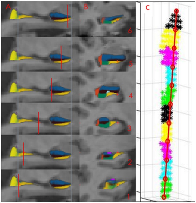
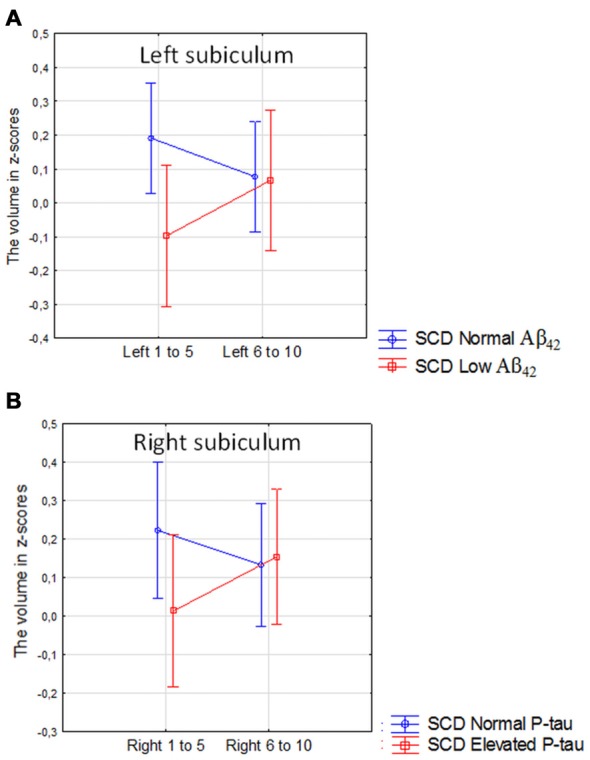
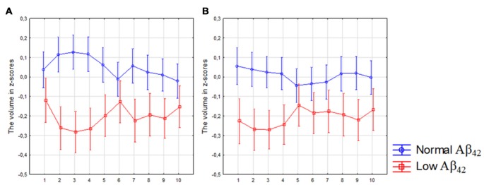
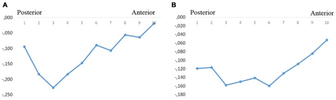
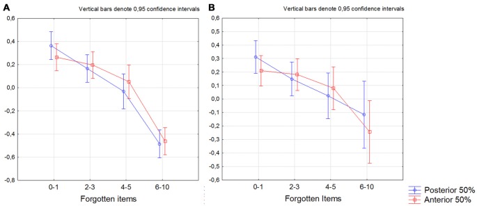
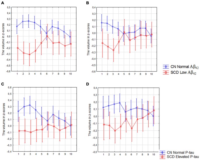
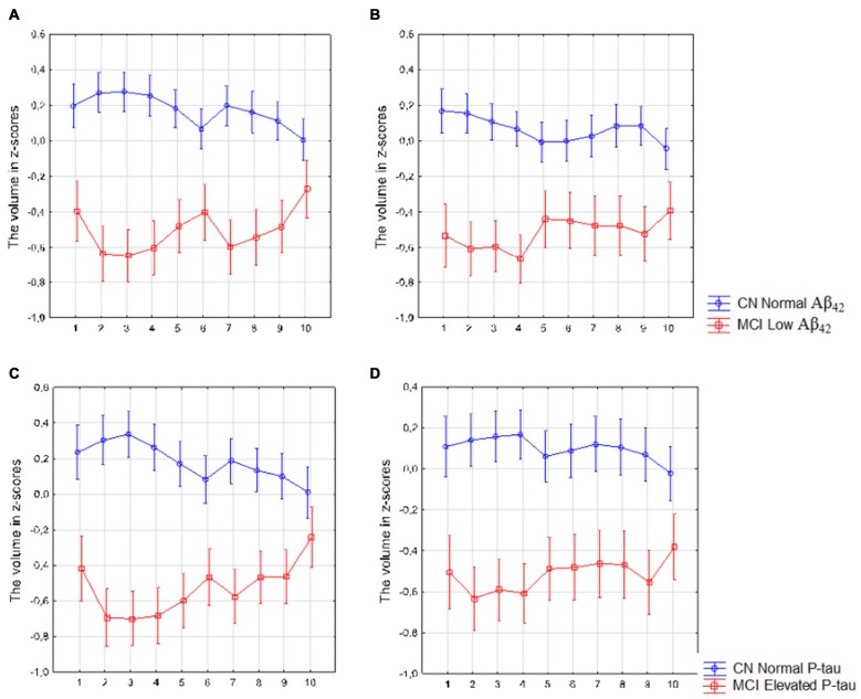
Alzheimer's disease (AD) is associated with atrophy of the cornu ammonis (CA) 1 and the subiculum subfield of the hippocampus (HC), and with deficits in episodic memory and spatial orientation. These deficits are mainly associated with the functionality of the posterior HC. We therefore hypothesized that key AD pathologies, i.e., β-amyloid and tau pathology would be particularly associated with the volume of the posterior subiculum in non-demented individuals. In our study we included 302 cognitively normal elderly participants (CN), 183 patients with subjective cognitive decline (SCD) and 171 patients with amnestic mild cognitive impairment (MCI), all of whom underwent 3T magnetic resonance images (MRI). The subicular subfield was segmented using Freesurfer 5.3 and divided into 10 volumetric segments moving from the most posterior (segment 1) to the most anterior part along the axis of the hippocampal head and body (segment 10). Cerebrospinal fluid (CSF) Aβ and phosphorylated tau (P-tau) were quantified using ELISA and were used as biomarkers for β-amyloid and tau pathology, respectively. In the total sample, tau-pathology and Aβ-pathology and (measured by elevated P-tau and low Aβ levels in CSF) and mild memory dysfunction were mostly associated with the volume changes of the posterior subiculum. Both SCD and MCI patients with elevated P-tau or low Aβ levels displayed predominantly posterior subicular atrophy in comparisons to control subjects with normal CSF biomarker levels. Finally, there was no main effect of Aβ or P-tau when comparing SCD with abnormal P-tau or Aβ with SCD with normal levels of these CSF-biomarkers. However, in the left subiculum there was a significant interaction revealing atrophy in the left posterior but not the anterior subiculum in participants with low Aβ levels. The same pattern was observed on the contralateral side in participants with elevated P-tau levels. In conclusion, AD pathologies and mild memory dysfunction are mainly associated with atrophy of the posterior parts of the subicular subfields of the HC in non-demented individuals. In light of these findings we suggest that segmentation of the HC subfields may benefit from considering the volume of the different anterior-posterior subsections of each subfield.
阿尔茨海默病(AD)与海马体(HC)的海马角(CA)1和下托亚区萎缩相关,且与情景记忆和空间定向缺陷有关。这些缺陷主要与后海马体的功能有关。因此,我们假设关键的AD病理特征,即β-淀粉样蛋白和tau病理特征,在非痴呆个体中会特别与后下托的体积相关。在我们的研究中,纳入了302名认知正常的老年参与者(CN)、183名主观认知下降(SCD)患者和171名遗忘型轻度认知障碍(MCI)患者,他们均接受了3T磁共振成像(MRI)检查。使用Freesurfer 5.3对下托亚区进行分割,并沿海马头部和体部的轴线从最后部(第1段)到最前部(第10段)将其分为10个体积段。使用酶联免疫吸附测定法(ELISA)对脑脊液(CSF)中的Aβ和磷酸化tau(P-tau)进行定量,分别将其用作β-淀粉样蛋白和tau病理特征的生物标志物。在整个样本中,tau病理特征和Aβ病理特征(通过脑脊液中P-tau升高和Aβ水平降低来衡量)以及轻度记忆功能障碍主要与后下托的体积变化相关。与脑脊液生物标志物水平正常的对照受试者相比,P-tau升高或Aβ水平降低的SCD和MCI患者均主要表现为后下托萎缩。最后,将脑脊液生物标志物水平异常的P-tau的SCD与正常水平的Aβ的SCD进行比较时,Aβ或P-tau没有主要影响。然而,在左侧下托中,存在显著的相互作用,显示Aβ水平低的参与者左侧后下托而非前下托出现萎缩。在P-tau水平升高的参与者的对侧也观察到相同的模式。总之,在非痴呆个体中,AD病理特征和轻度记忆功能障碍主要与海马体下托亚区后部萎缩相关。鉴于这些发现,我们建议海马体亚区的分割可能受益于考虑每个亚区不同前后节段的体积。