Clínica Josefina Arregui, Alsasua, Spain.
Instituto de Neurociencias de Alicante (UMH-CSIC), Alicante, Spain.
Alzheimers Res Ther. 2022 Jul 22;14(1):98. doi: 10.1186/s13195-022-01031-6.
People with subjective cognitive decline (SCD) report cognitive deterioration. However, their performance in neuropsychological evaluation falls within the normal range. The present study aims to analyse whether structural magnetic resonance imaging (MRI) reveals grey matter changes in the SCD population compared with healthy normal controls (HC).
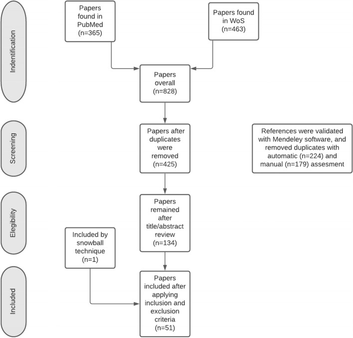
Parallel systematic searches in PubMed and Web of Science databases were conducted, following the Preferred Reporting Items for Systematic Reviews and Meta-Analyses (PRISMA) guidelines. Quality assessment was completed using the Newcastle-Ottawa Scale (NOS).
Fifty-one MRI studies were included. Thirty-five studies used a region of interest (ROI) analysis, 15 used a voxel-based morphometry (VBM) analysis and 10 studies used a cortical thickness (CTh) analysis. Ten studies combined both, VBM or CTh analysis with ROI analysis.
Medial temporal structures, like the hippocampus or the entorhinal cortex (EC), seemed to present grey matter reduction in SCD compared with HC, but the samples and results are heterogeneous. Larger sample sizes could help to better determine if these grey matter changes are consistent in SCD subjects.
有主观认知下降(SCD)的人报告认知能力下降。然而,他们在神经心理学评估中的表现仍在正常范围内。本研究旨在分析与健康正常对照组(HC)相比,结构性磁共振成像(MRI)是否显示 SCD 人群的灰质变化。
按照系统评价和荟萃分析的首选报告项目(PRISMA)指南,在 PubMed 和 Web of Science 数据库中进行平行系统搜索。使用纽卡斯尔-渥太华量表(NOS)进行质量评估。
共纳入 51 项 MRI 研究。35 项研究使用了感兴趣区(ROI)分析,15 项研究使用了基于体素的形态测量学(VBM)分析,10 项研究使用了皮质厚度(CTh)分析。10 项研究将 VBM 或 CTh 分析与 ROI 分析相结合。
与 HC 相比,SCD 中内侧颞叶结构,如海马体或内嗅皮层(EC),似乎存在灰质减少,但样本和结果存在异质性。更大的样本量可能有助于更好地确定 SCD 患者的这些灰质变化是否一致。