Group of Radiation Biology and Tumor Physiology, Department of Radiation Biology, Institute for Cancer Research, Oslo University Hospital, Oslo, Norway.
Department of Radiation Biology, Institute for Cancer Research, Norwegian Radium Hospital, Box 4953, Nydalen, 0424, Oslo, Norway.
J Transl Med. 2017 Oct 10;15(1):203. doi: 10.1186/s12967-017-1307-4.
Malignant melanoma of the skin can metastasize through blood vessels and lymphatics. The primary tumor develops a vascular microenvironment characterized by abnormal blood vessels and lymphatics and a physicochemical microenvironment characterized by low oxygen tension, regions with hypoxic tissue, and high interstitial fluid pressure (IFP). This study aimed at identifying relationships between the metastatic route of melanomas and characteristic features of the microvascular and physicochemical microenvironments of the primary tumor.
Two patient-derived xenograft (PDX) models (E-13, N-15) and four cell line-derived xenografts (CDX) models (C-10, D-12, R-18, T-22) of human melanoma were included in the study. Tumors were transplanted to an orthotopic site in BALB/c-nu/nu mice, and when the tumors had grown to a volume of 500-600 mm, the IFP of the primary tumor was measured and the hypoxia marker pimonidazole was administered before the host mouse was euthanized. The primary tumor, lungs, and six pairs of lymph nodes were evaluated by examining hematoxylin/eosin-stained and immunostained histological preparations. The expression of angiogenesis-related genes was assessed by quantitative PCR.
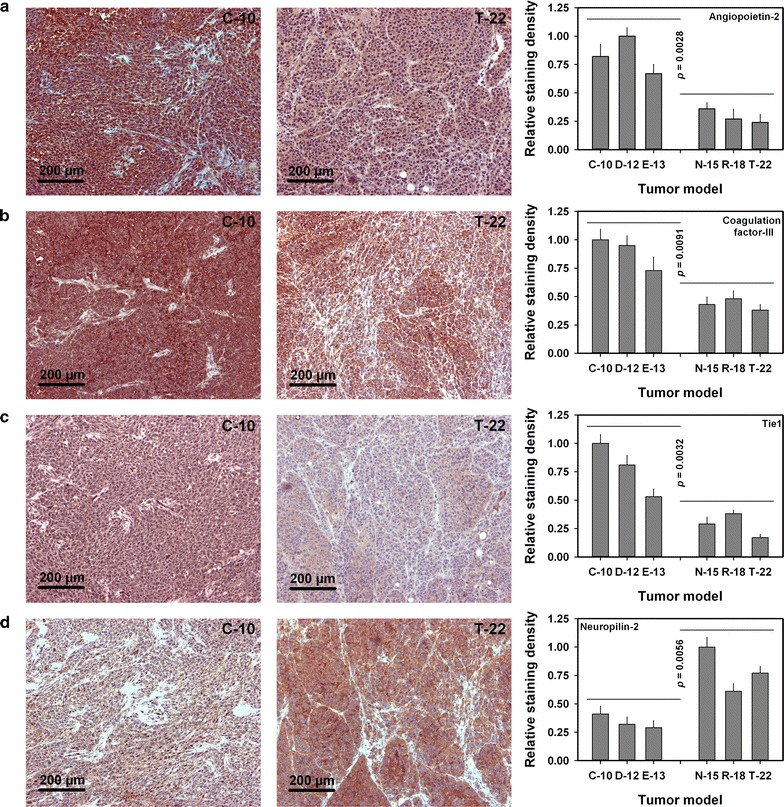
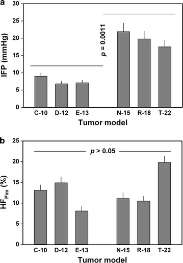
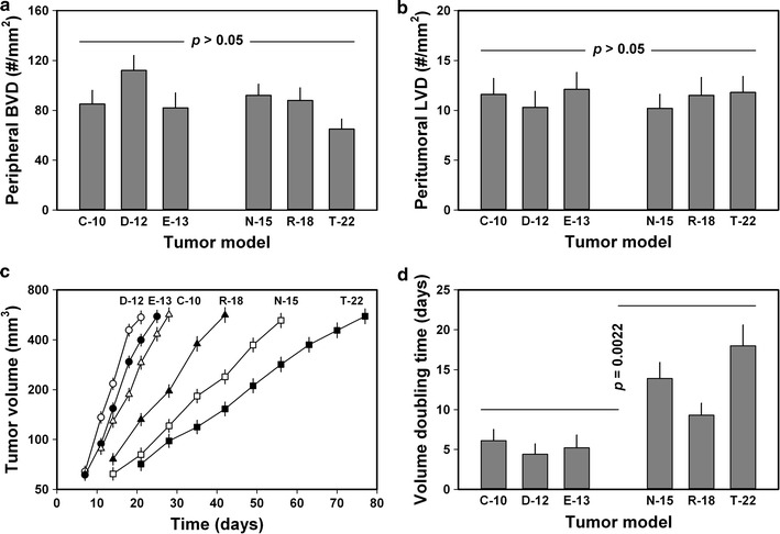
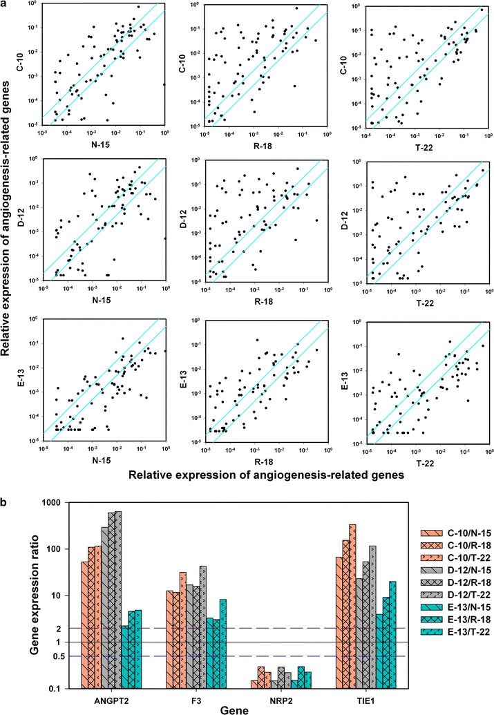
C-10, D-12, and E-13 tumors disseminated primarily by the hematogenous route and developed pulmonary metastases. These tumors showed high angiogenic activity and high expression of the F3 gene as well as ANGPT2 and TIE1, genes encoding proteins of the angiopoietin-tie system. N-15, R-18, and T-22 tumors disseminated mainly by the lymphogenous route and developed metastases in draining lymph nodes. These tumors had highly elevated IFP and showed high expression of NRP2, a gene encoding neuropilin-2.
The primary metastatic route of orthotopic human melanoma xenografts and the development of lung and lymph node metastases are influenced significantly by the microvascular and physicochemical microenvironments of the primary tumor.
皮肤恶性黑色素瘤可通过血管和淋巴管转移。原发肿瘤形成的血管微环境具有特征性,包括异常的血管和淋巴管;理化微环境具有特征性,包括低氧张力、缺氧组织区域和高间质液压力(IFP)。本研究旨在确定黑色素瘤的转移途径与原发肿瘤微血管和理化微环境特征之间的关系。
本研究纳入了 2 个患者来源的异种移植(PDX)模型(E-13、N-15)和 4 个细胞系来源的异种移植(CDX)模型(C-10、D-12、R-18、T-22)。肿瘤被移植到 BALB/c-nu/nu 小鼠的原位,当肿瘤生长到 500-600mm3 时,测量原发肿瘤的 IFP,并在处死宿主小鼠之前给予缺氧标记物 pimonidazole。通过检查苏木精/伊红染色和免疫组织化学染色的组织学切片评估原发肿瘤、肺和 6 对淋巴结。通过定量 PCR 评估与血管生成相关的基因表达。
C-10、D-12 和 E-13 肿瘤主要通过血行途径扩散,并发展为肺转移。这些肿瘤表现出高度的血管生成活性和 F3 基因以及 ANGPT2 和 TIE1(编码血管生成素- tie 系统蛋白的基因)的高表达。N-15、R-18 和 T-22 肿瘤主要通过淋巴途径扩散,并在引流淋巴结中发展转移。这些肿瘤的 IFP 显著升高,并且 NRP2(编码神经纤毛蛋白-2 的基因)表达水平较高。
原位人黑色素瘤异种移植的主要转移途径以及肺和淋巴结转移的发生,受原发肿瘤微血管和理化微环境的显著影响。