Han Han, Zhou Hui, Li Jing, Feng Xiuyan, Zou Dan, Zhou Weiqiang
Key Laboratory of Environmental Pollution and Microecology of Liaoning Province, Shenyang Medical College, No.146 North Huanghe St, Huanggu Dis, Shenyang City, Liaoning Pro 110034, China.
Department of Biochemistry and Molecular Biology, Shenyang Medical College, No.146 North Huanghe St, Huanggu Dis, Shenyang City, Liaoning Pro 110034, China.
Cell Death Discov. 2017 Aug 21;3:17052. doi: 10.1038/cddiscovery.2017.52. eCollection 2017.
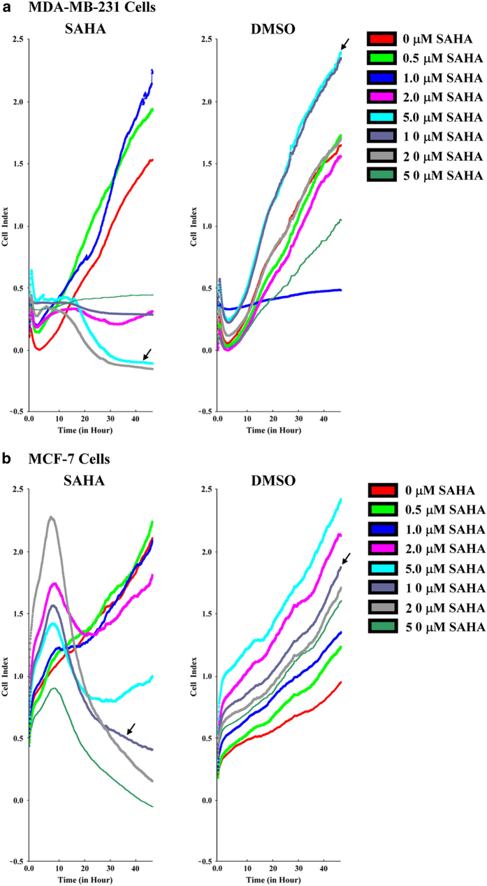
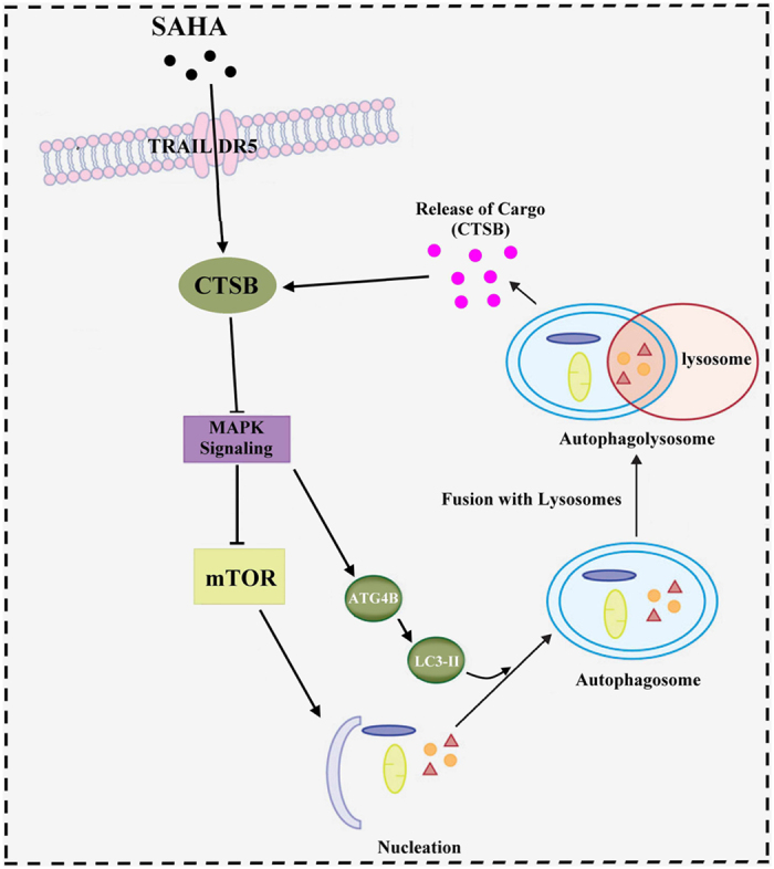
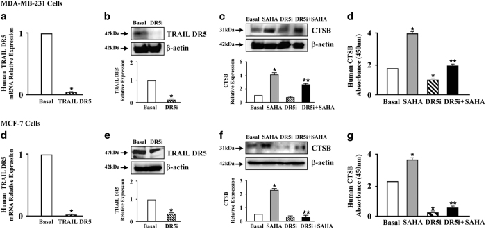
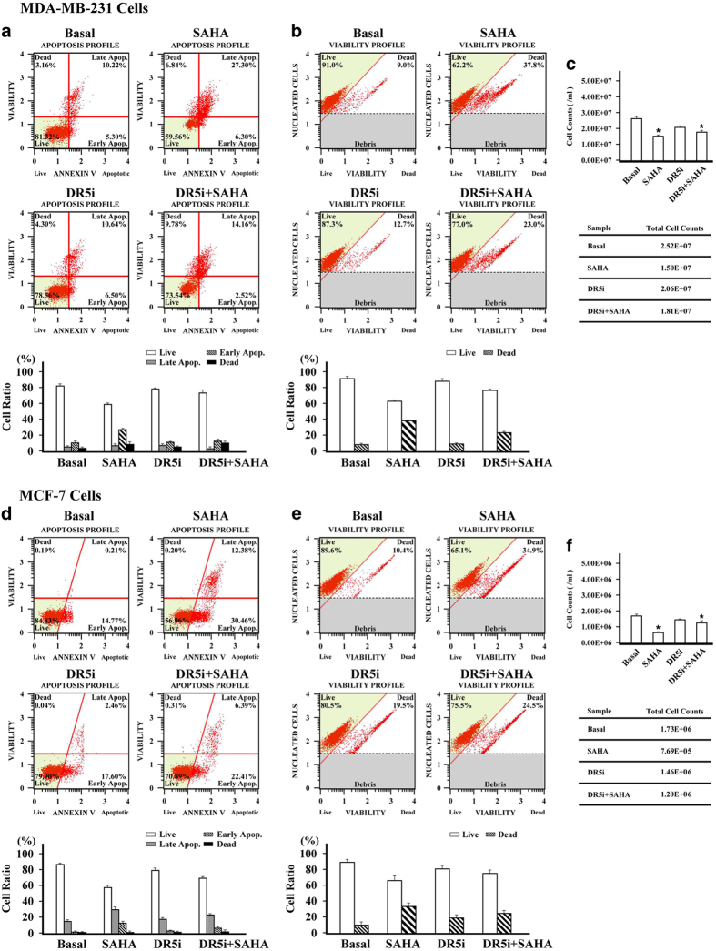
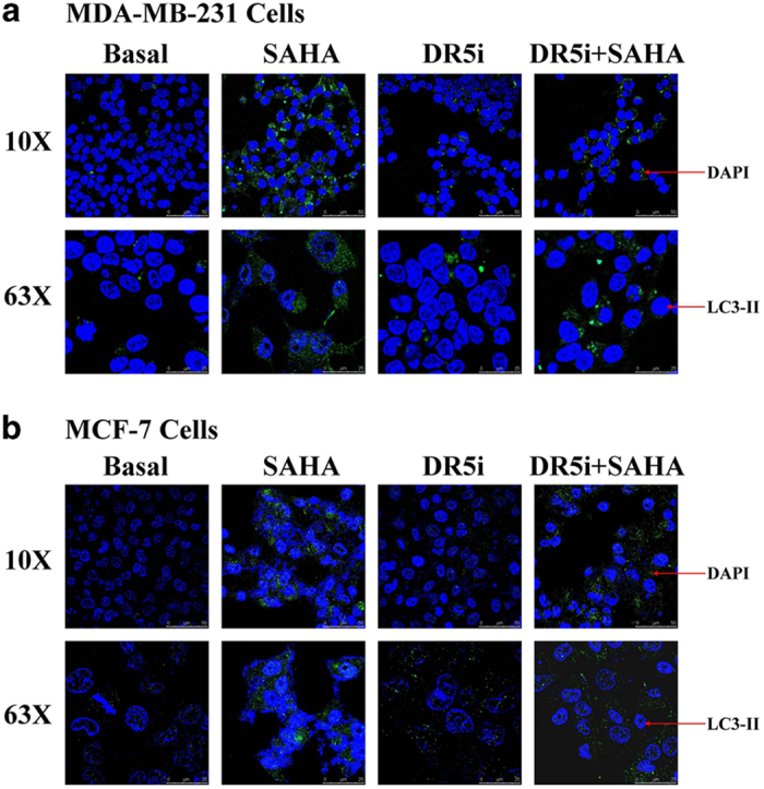
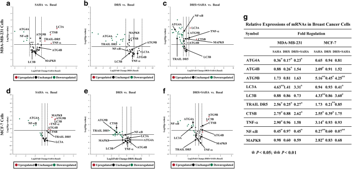
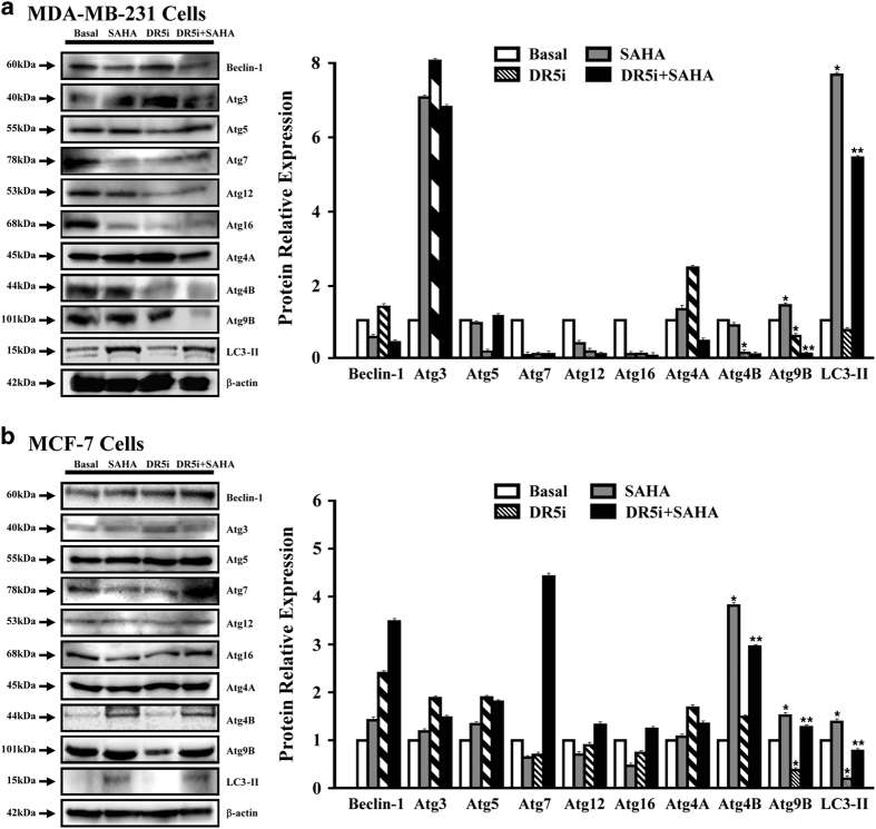
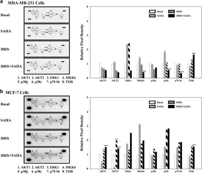
To investigate the ability of SAHA-induced TRAIL DR5-CTSB crosstalk to initiate the breast cancer autophagy, RTCA assay was performed to assess the effect of SAHA on breast cancer cells, and western blot and ELISA were used to verify the inductive effects on expression of CTSB. Breast cancer cells were transfected with TRAIL DR5 siRNA to block the function of TRAIL DR5. Cell viability and apoptosis of breast cancer cells were analyzed using a muse cell analyzer. The distribution of LC3-II in TRAIL DR5-silenced breast cancer cells treated with SAHA was observed by immunofluorescence microscopy, the mRNA levels of autophagy-related genes were detected by RNA microarray, and the activity of autophagy-related signaling pathways was screened by MAPK antibody array. Results indicated that SAHA did indeed repress the growth of breast cancer cell lines with inducing CTSB expression. Western blot and ELISA results indicated that TRAIL DR5 was involved in the expression of CTSB in SAHA-induced breast cancer cells. Cell viability and apoptosis assays showed that the inactivation of TRAIL DR5 can significantly inhibit the effects of SAHA. An immunofluorescence assay indicated that, with SAHA treatment, MDA-MB-231 and MCF-7 cells underwent apparent morphological changes. While SAHA was added in the TRAIL-DR5 blocked cells, the distribution of LC3-II signal was dispersed, the intensity of fluorescence signal was weaker than that of SAHA alone. RNA array indicated that SAHA significantly increased mRNA expression of autophagy marker LC3A/B whereas the change was significantly reversed in TRAIL DR5-silenced cells. The results of MAPK antibody array showed that SAHA and TRAIL DR5 could affect the activity of AKT1, AKT2, and TOR protein in breast cancer cells. These results provide more evidence that SAHA may stimulate TRAIL DR5-CTSB crosstalk, influence the activity of downstream TOR signalling pathway mainly through the AKTs pathway, and initiate the autophagy of breast cancer cells.
为了研究SAHA诱导的TRAIL DR5-CTSB相互作用引发乳腺癌自噬的能力,进行了实时无标记细胞分析(RTCA)以评估SAHA对乳腺癌细胞的影响,并使用蛋白质免疫印迹法和酶联免疫吸附测定(ELISA)来验证其对组织蛋白酶B(CTSB)表达的诱导作用。用TRAIL DR5小干扰RNA(siRNA)转染乳腺癌细胞以阻断TRAIL DR5的功能。使用缪斯细胞分析仪分析乳腺癌细胞的活力和凋亡情况。通过免疫荧光显微镜观察SAHA处理的TRAIL DR5沉默乳腺癌细胞中微管相关蛋白1轻链3-II(LC3-II)的分布,通过RNA微阵列检测自噬相关基因的mRNA水平,并通过丝裂原活化蛋白激酶(MAPK)抗体阵列筛选自噬相关信号通路的活性。结果表明,SAHA确实通过诱导CTSB表达来抑制乳腺癌细胞系的生长。蛋白质免疫印迹法和ELISA结果表明,TRAIL DR5参与了SAHA诱导的乳腺癌细胞中CTSB的表达。细胞活力和凋亡分析表明,TRAIL DR5失活可显著抑制SAHA的作用。免疫荧光分析表明,经SAHA处理后,MDA-MB-231和MCF-7细胞发生明显的形态变化。当在TRAIL-DR5阻断的细胞中加入SAHA时,LC3-II信号的分布分散,荧光信号强度比单独使用SAHA时弱。RNA阵列表明,SAHA显著增加自噬标志物LC3A/B的mRNA表达,而在TRAIL DR5沉默的细胞中这种变化显著逆转。MAPK抗体阵列的结果表明,SAHA和TRAIL DR5可影响乳腺癌细胞中蛋白激酶B1(AKT1)、蛋白激酶B2(AKT2)和雷帕霉素靶蛋白(TOR)的活性。这些结果提供了更多证据,表明SAHA可能刺激TRAIL DR5-CTSB相互作用,主要通过AKT信号通路影响下游TOR信号通路的活性,并引发乳腺癌细胞的自噬。