Le Roux Pierre-Yves, Siva Shankar, Callahan Jason, Claudic Yannis, Bourhis David, Steinfort Daniel P, Hicks Rodney J, Hofman Michael S
Cancer Imaging, Peter MacCallum Cancer Centre, 305 Grattan St, Melbourne, 3000, Australia.
Nuclear Medicine Department, Brest University Hospital, EA3878 (GETBO) IFR, 148, Brest, France.
EJNMMI Res. 2017 Oct 10;7(1):82. doi: 10.1186/s13550-017-0332-x.
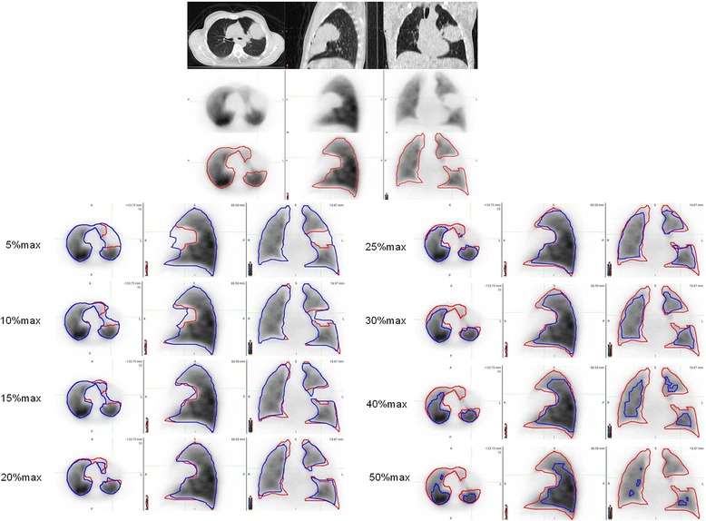
Functional volumes computed from Ga-ventilation/perfusion (V/Q) PET/CT, which we have shown to correlate with pulmonary function test parameters (PFTs), have potential diagnostic utility in a variety of clinical applications, including radiotherapy planning. An automatic segmentation method would facilitate delineation of such volumes. The aim of this study was to develop an automated threshold-based approach to delineate functional volumes that best correlates with manual delineation. Thirty lung cancer patients undergoing both V/Q PET/CT and PFTs were analyzed. Images were acquired following inhalation of Galligas and, subsequently, intravenous administration of Ga-macroaggreted-albumin (MAA). Using visually defined manual contours as the reference standard, various cutoff values, expressed as a percentage of the maximal pixel value, were applied. The average volume difference and Dice similarity coefficient (DSC) were calculated, measuring the similarity of the automatic segmentation and the reference standard. Pearson's correlation was also calculated to compare automated volumes with manual volumes, and automated volumes optimized to PFT indices.
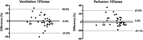
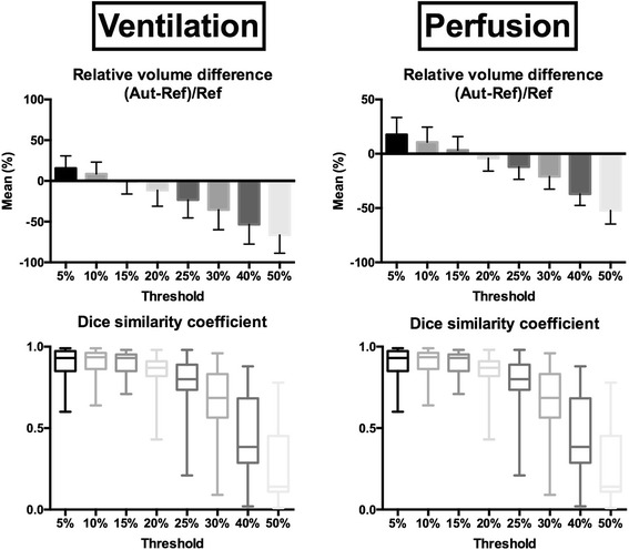
For ventilation volumes, mean volume difference was lowest (- 0.4%) using a 15%max threshold with Pearson's coefficient of 0.71. Applying this cutoff, median DSC was 0.93 (0.87-0.95). Nevertheless, limits of agreement in volume differences were large (- 31.0 and 30.2%) with differences ranging from - 40.4 to + 33.0%. For perfusion volumes, mean volume difference was lowest and Pearson's coefficient was highest using a 15%max threshold (3.3% and 0.81, respectively). Applying this cutoff, median DSC was 0.93 (0.88-0.93). Nevertheless, limits of agreement were again large (- 21.1 and 27.8%) with volume differences ranging from - 18.6 to + 35.5%. Using the 15%max threshold, moderate correlation was demonstrated with FEV1/FVC (r = 0.48 and r = 0.46 for ventilation and perfusion images, respectively). No correlation was found between other PFT indices.
To automatically delineate functional volumes with Ga-V/Q PET/CT, the most appropriate cutoff was 15%max for both ventilation and perfusion images. However, using this unique threshold systematically provided unacceptable variability compared to the reference volume and relatively poor correlation with PFT parameters. Accordingly, a visually adapted semi-automatic method is favored, enabling rapid and quantitative delineation of lung functional volumes with Ga-V/Q PET/CT.
通过镓通气/灌注(V/Q)PET/CT计算得出的功能体积,我们已证明其与肺功能测试参数(PFTs)相关,在包括放射治疗计划在内的各种临床应用中具有潜在的诊断效用。一种自动分割方法将有助于描绘此类体积。本研究的目的是开发一种基于阈值的自动方法来描绘与手动描绘最相关的功能体积。对30例同时接受V/Q PET/CT和PFTs检查的肺癌患者进行了分析。在吸入镓气体后,随后静脉注射镓标记的大聚合白蛋白(MAA)后采集图像。以视觉定义的手动轮廓作为参考标准,应用了各种截止值,以最大像素值的百分比表示。计算了平均体积差异和骰子相似系数(DSC),以测量自动分割与参考标准的相似性。还计算了皮尔逊相关性,以比较自动体积与手动体积,以及根据PFT指标优化的自动体积。
对于通气体积,使用15%max阈值时平均体积差异最低(-0.4%),皮尔逊系数为0.71。应用此截止值时,中位数DSC为0.93(0.87 - 0.95)。然而,体积差异的一致性界限很大(-31.0%和30.2%),差异范围为-40.4%至+33.0%。对于灌注体积,使用15%max阈值时平均体积差异最低,皮尔逊系数最高(分别为3.3%和0.81)。应用此截止值时,中位数DSC为0.93(0.88 - 0.93)。然而,一致性界限再次很大(-21.1%和27.8%),体积差异范围为-18.6%至+35.5%。使用15%max阈值时,与FEV1/FVC显示出中等相关性(通气和灌注图像的r分别为0.48和0.46)。未发现与其他PFT指标之间存在相关性。
为了通过镓V/Q PET/CT自动描绘功能体积,通气和灌注图像最合适的截止值均为15%max。然而,与参考体积相比,使用这个单一阈值系统地产生了不可接受的变异性,并且与PFT参数的相关性相对较差。因此,更倾向于一种视觉适应的半自动方法,它能够通过镓V/Q PET/CT快速且定量地描绘肺功能体积。