McIntosh Lachlan, Jackson Price, Hardcastle Nicholas, Bressel Mathias, Kron Tomas, Callahan Jason W, Steinfort Daniel, Bucknell Nicholas, Hofman Michael S, Siva Shankar
Department of Physical Sciences, Peter MacCallum Cancer Centre, Melbourne, 3000, Australia.
Department of Cancer Imaging, Peter MacCallum Cancer Centre, Melbourne, 3000, Australia.
EJNMMI Phys. 2021 Mar 7;8(1):23. doi: 10.1186/s40658-021-00375-6.
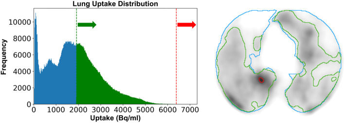
Functional lung mapping from Ga-ventilation/perfusion (V/Q) PET/CT, which has been shown to correlate with pulmonary function tests (PFTs), may be beneficial in a number of clinical applications where sparing regions of high lung function is of interest. Regions of clumping in the proximal airways in patients with airways disease can result in areas of focal intense activity and artefact in ventilation imaging. These artefacts may even shine through to subsequent perfusion images and create a challenge for quantitative analysis of PET imaging. We aimed to develop an automated algorithm that interprets the uptake histogram of PET images to calculate a peak uptake value more representative of the global lung volume.
Sixty-six patients recruited from a prospective clinical trial underwent both V/Q PET/CT imaging and PFT analysis before treatment. PET images were normalised using an iterative histogram analysis technique to account for tracer hotspots prior to the threshold-based delineation of varying values. Pearson's correlation between fractional lung function and PFT score was calculated for ventilation, perfusion, and matched imaging volumes at varying threshold values.
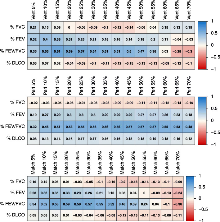
For all functional imaging thresholds, only FEV1/FVC PFT yielded reasonable correlations to image-based functional volume. For ventilation, a range of 10-30% of adapted peak uptake value provided a reasonable threshold to define a volume that correlated with FEV1/FVC (r = 0.54-0.61). For perfusion imaging, a similar correlation was observed (r = 0.51-0.56) in the range of 20-60% adapted peak threshold. Matched volumes were closely linked to ventilation with a threshold range of 15-35% yielding a similar correlation (r = 0.55-0.58).
Histogram normalisation may be implemented to determine the presence of tracer clumping hotspots in Ga-68 V/Q PET imaging allowing for automated delineation of functional lung and standardisation of functional volume reporting.
Ga通气/灌注(V/Q)PET/CT的功能性肺成像已被证明与肺功能测试(PFT)相关,在许多关注保留高肺功能区域的临床应用中可能有益。气道疾病患者近端气道中的聚集区域可导致通气成像中出现局灶性高强度活动区域和伪影。这些伪影甚至可能透射到后续的灌注图像中,给PET成像的定量分析带来挑战。我们旨在开发一种自动算法,通过解释PET图像的摄取直方图来计算更能代表全肺容积的峰值摄取值。
从一项前瞻性临床试验中招募的66名患者在治疗前接受了V/Q PET/CT成像和PFT分析。在基于阈值描绘不同值之前,使用迭代直方图分析技术对PET图像进行归一化,以考虑示踪剂热点。计算不同阈值下通气、灌注和匹配成像容积的肺功能分数与PFT评分之间的Pearson相关性。
对于所有功能成像阈值,只有FEV1/FVC PFT与基于图像的功能容积有合理的相关性。对于通气,调整后的峰值摄取值的10%-30%范围提供了一个合理的阈值来定义与FEV1/FVC相关的容积(r = 0.54-0.61)。对于灌注成像,在调整后的峰值阈值的20%-60%范围内观察到类似的相关性(r = 0.51-0.56)。匹配容积与通气密切相关,阈值范围为15%-35%时产生类似的相关性(r = 0.55-0.58)。
可实施直方图归一化以确定Ga-68 V/Q PET成像中示踪剂聚集热点的存在,从而实现功能性肺的自动描绘和功能容积报告的标准化。