• 文献检索
  • 文档翻译
  • 深度研究
  • 学术资讯
  • Suppr Zotero 插件Zotero 插件
  • 邀请有礼
  • 套餐&价格
  • 历史记录
应用&插件
Suppr Zotero 插件Zotero 插件浏览器插件Mac 客户端Windows 客户端微信小程序
定价
高级版会员购买积分包购买API积分包
服务
文献检索文档翻译深度研究API 文档MCP 服务
关于我们
关于 Suppr公司介绍联系我们用户协议隐私条款
关注我们

Suppr 超能文献

核心技术专利:CN118964589B侵权必究
粤ICP备2023148730 号-1Suppr @ 2026

文献检索

告别复杂PubMed语法,用中文像聊天一样搜索,搜遍4000万医学文献。AI智能推荐,让科研检索更轻松。

立即免费搜索

文件翻译

保留排版,准确专业,支持PDF/Word/PPT等文件格式,支持 12+语言互译。

免费翻译文档

深度研究

AI帮你快速写综述,25分钟生成高质量综述,智能提取关键信息,辅助科研写作。

立即免费体验

脊髓延髓肌萎缩症患者吞咽功能障碍的定量评估

Quantitative Assessment of Swallowing Dysfunction in Patients with Spinal and Bulbar Muscular Atrophy.

作者信息

Hashizume Atsushi, Banno Haruhiko, Katsuno Masahisa, Hijikata Yasuhiro, Yamada Shinichiro, Inagaki Tomonori, Suzuki Keisuke, Sobue Gen

机构信息

Department of Neurology, Nagoya University Graduate School of Medicine, Japan.

Department of Clinical Research, Innovation Center for Clinical Research, National Center for Geriatrics and Gerontology, Japan.

出版信息

Intern Med. 2017 Dec 1;56(23):3159-3165. doi: 10.2169/internalmedicine.8799-16. Epub 2017 Oct 11.

DOI:10.2169/internalmedicine.8799-16
PMID:29021456
原文链接:https://pmc.ncbi.nlm.nih.gov/articles/PMC5742386/
Abstract

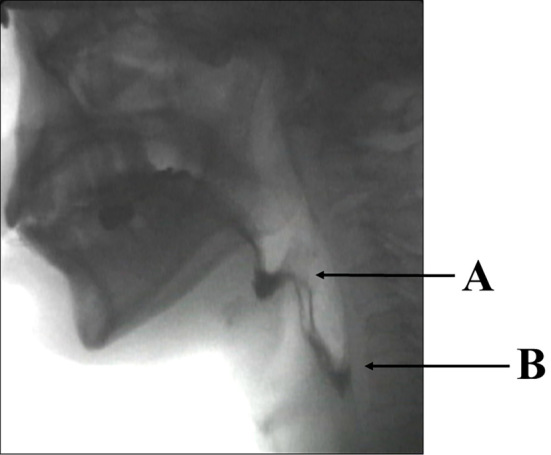
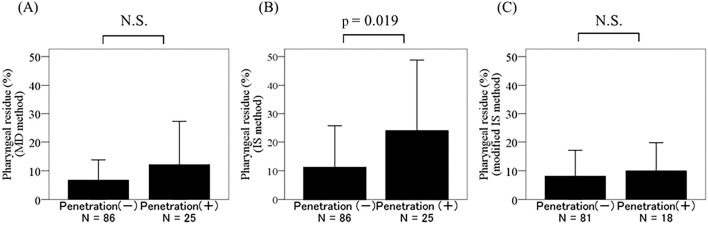
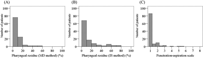
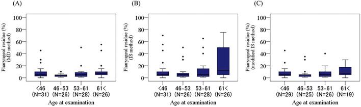
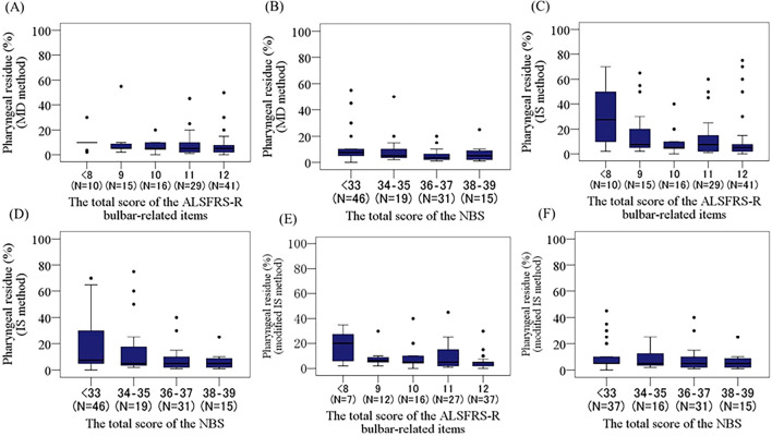
Objective This study aimed to evaluate swallowing dysfunction in patients with spinal and bulbar muscular atrophy and to identify the most appropriate method of assessing swallowing dysfunction using a videofluoroscopic swallowing study. Methods In the videofluoroscopic swallowing study, patients were instructed to swallow 3 mL of 40% weight/volume barium sulfate twice, and the pharyngeal residue was measured. We used three different methods to quantify the pharyngeal barium residue and an eight-point scale to evaluate the laryngeal penetration leading to aspiration pneumoniae. Patients We assessed 111 patients with spinal and bulbar muscular atrophy who weren't undergoing disease-specific treatment. Results Our results showed that the pharyngeal barium residue after initial swallowing correlated better with the bulbar-related functional rating scales than that after multiple deglutition. This correlation was vague when the data from patients whose barium residue was >50% were eliminated. In addition, evaluating the pharyngeal residue after initial swallowing proved to be the most sensitive method with regard to laryngeal penetration. Conclusion This study showed that the pharyngeal barium residue after initial swallowing was the most appropriate parameter for quantitatively assessing the degree of dysphagia using a videofluoroscopic swallowing study and suggests that this method may predict laryngeal penetration and aspiration in patients with spinal and bulbar muscular atrophy.

摘要

目的 本研究旨在评估脊髓延髓肌肉萎缩症患者的吞咽功能障碍,并通过电视荧光吞咽造影研究确定评估吞咽功能障碍的最合适方法。方法 在电视荧光吞咽造影研究中,指导患者分两次吞咽3 mL 40%重量/体积的硫酸钡,并测量咽部残留量。我们使用三种不同方法对咽部钡剂残留进行量化,并使用八分制量表评估导致吸入性肺炎的喉穿透情况。患者 我们评估了111例未接受疾病特异性治疗的脊髓延髓肌肉萎缩症患者。结果 我们的结果表明,初次吞咽后的咽部钡剂残留与延髓相关功能评定量表的相关性优于多次吞咽后。当排除钡剂残留>50%的患者数据时,这种相关性不明确。此外,就喉穿透而言,评估初次吞咽后的咽部残留是最敏感的方法。结论 本研究表明,初次吞咽后的咽部钡剂残留是使用电视荧光吞咽造影研究定量评估吞咽困难程度的最合适参数,并表明该方法可能预测脊髓延髓肌肉萎缩症患者的喉穿透和误吸情况。

https://cdn.ncbi.nlm.nih.gov/pmc/blobs/9e91/5742386/0bf612b0f7eb/1349-7235-56-3159-g005.jpg
https://cdn.ncbi.nlm.nih.gov/pmc/blobs/9e91/5742386/9e4f68788c99/1349-7235-56-3159-g001.jpg
https://cdn.ncbi.nlm.nih.gov/pmc/blobs/9e91/5742386/f3cec3edbfce/1349-7235-56-3159-g002.jpg
https://cdn.ncbi.nlm.nih.gov/pmc/blobs/9e91/5742386/806a5c32c4df/1349-7235-56-3159-g003.jpg
https://cdn.ncbi.nlm.nih.gov/pmc/blobs/9e91/5742386/2ef520be30d1/1349-7235-56-3159-g004.jpg
https://cdn.ncbi.nlm.nih.gov/pmc/blobs/9e91/5742386/0bf612b0f7eb/1349-7235-56-3159-g005.jpg

相似文献

1
Quantitative Assessment of Swallowing Dysfunction in Patients with Spinal and Bulbar Muscular Atrophy.脊髓延髓肌萎缩症患者吞咽功能障碍的定量评估
Intern Med. 2017 Dec 1;56(23):3159-3165. doi: 10.2169/internalmedicine.8799-16. Epub 2017 Oct 11.
2
Swallowing markers in spinal and bulbar muscular atrophy.脊髓延髓肌肉萎缩症中的吞咽标志物
Ann Clin Transl Neurol. 2017 May 24;4(8):534-543. doi: 10.1002/acn3.425. eCollection 2017 Aug.
3
Effect of leuprorelin in bulbar function of spinal and bulbar muscular atrophy patients: observational study for 1 year.促性腺激素释放激素对延髓和球部脊髓性肌萎缩症患者球部功能的影响:为期 1 年的观察性研究。
J Neurol. 2021 Sep;268(9):3344-3351. doi: 10.1007/s00415-021-10503-y. Epub 2021 Mar 6.
4
A Preliminary Videofluoroscopic Investigation of Swallowing Physiology and Function in Individuals with Oculopharyngeal Muscular Dystrophy (OPMD).眼咽型肌营养不良症(OPMD)患者吞咽生理与功能的初步视频荧光造影研究
Dysphagia. 2018 Dec;33(6):789-802. doi: 10.1007/s00455-018-9904-9. Epub 2018 May 3.
5
The modified barium swallow and the functional endoscopic evaluation of swallowing.改良钡餐吞咽造影和吞咽功能内镜评估
Otolaryngol Clin North Am. 2013 Dec;46(6):1009-22. doi: 10.1016/j.otc.2013.08.001. Epub 2013 Oct 8.
6
Quantifying swallowing function after stroke: A functional dysphagia scale based on videofluoroscopic studies.量化中风后的吞咽功能:基于视频荧光透视研究的功能性吞咽困难量表
Arch Phys Med Rehabil. 2001 May;82(5):677-82. doi: 10.1053/apmr.2001.21939.
7
Efficacy and safety of leuprorelin in patients with spinal and bulbar muscular atrophy (JASMITT study): a multicentre, randomised, double-blind, placebo-controlled trial.在脊髓性肌萎缩症(JASMITT 研究)患者中使用亮丙瑞林的疗效和安全性:一项多中心、随机、双盲、安慰剂对照试验。
Lancet Neurol. 2010 Sep;9(9):875-84. doi: 10.1016/S1474-4422(10)70182-4. Epub 2010 Aug 4.
8
Swallowing Problems in Spinal Muscular Atrophy Types 2 and 3: A Clinical, Videofluoroscopic and Ultrasound Study.脊髓性肌萎缩症 2 型和 3 型的吞咽问题:一项临床、视频透视和超声研究。
J Neuromuscul Dis. 2023;10(3):427-438. doi: 10.3233/JND-221640.
9
Dysphagia in X-linked bulbospinal muscular atrophy (Kennedy disease).X连锁性球脊髓性肌萎缩症(肯尼迪病)中的吞咽困难
Neuromuscul Disord. 2009 Oct;19(10):704-8. doi: 10.1016/j.nmd.2009.06.371. Epub 2009 Jul 17.
10
Videofluoroscopic evaluation of pharyngeal swallowing dysfunction after esophagectomy with three-field lymph node dissection.食管癌切除联合三野淋巴结清扫术后咽吞咽功能障碍的电视荧光吞咽功能评估
Eur Arch Otorhinolaryngol. 2017 Jan;274(1):321-326. doi: 10.1007/s00405-016-4209-9. Epub 2016 Jul 16.

引用本文的文献

1
Rehabilitation of Post-Stroke Swallowing Dysfunction with Repeated Transcranial Magnetic Stimulation RTMS Based on Tomographic Images.基于断层图像的重复经颅磁刺激(rTMS)治疗脑卒中后吞咽功能障碍的康复。
Contrast Media Mol Imaging. 2022 Sep 13;2022:1118745. doi: 10.1155/2022/1118745. eCollection 2022.
2
The Role of Imaging Modalities in Diagnosing Dysphagia: A Clinical Review.影像学检查方法在吞咽困难诊断中的作用:一项临床综述
Cureus. 2021 Jul 31;13(7):e16786. doi: 10.7759/cureus.16786. eCollection 2021 Jul.
3
Dysphagia Phenotypes in Spinal Muscular Atrophy: The Past, Present, and Promise for the Future.

本文引用的文献

1
Two-dimensional perceptual videofluoroscopic swallowing analysis of the pharyngeal phase in patients older than 50 years.50岁以上患者咽期的二维感知性视频荧光吞咽分析
Arq Bras Cir Dig. 2013 Nov-Dec;26(4):274-9. doi: 10.1590/s0102-67202013000400005.
2
Tongue pressure as a novel biomarker of spinal and bulbar muscular atrophy.舌压作为脊髓性肌萎缩和球部肌萎缩的新型生物标志物。
Neurology. 2014 Jan 21;82(3):255-62. doi: 10.1212/WNL.0000000000000041. Epub 2013 Dec 18.
3
Videoendoscopic assessment of swallowing function to predict the future incidence of pneumonia of the elderly.
脊髓性肌萎缩症的吞咽障碍表型:过去、现在和未来的前景。
Am J Speech Lang Pathol. 2021 May 18;30(3):1008-1022. doi: 10.1044/2021_AJSLP-20-00217. Epub 2021 Apr 6.
4
Maximum bite force in patients with spinal muscular atrophy during the first year of nusinersen therapy - A pilot study.脊髓性肌萎缩症患者在接受nusinersen 治疗的第一年的最大咬合力 - 一项初步研究。
Acta Myol. 2020 Jun 1;39(2):83-89. doi: 10.36185/2532-1900-010. eCollection 2020 Jun.
5
Efficacy and safety of leuprorelin acetate for subjects with spinal and bulbar muscular atrophy: pooled analyses of two randomized-controlled trials.醋酸亮丙瑞林治疗脊髓性肌萎缩症和延髓性肌肉萎缩症受试者的疗效和安全性:两项随机对照试验的汇总分析。
J Neurol. 2019 May;266(5):1211-1221. doi: 10.1007/s00415-019-09251-x. Epub 2019 Mar 7.
6
Biomarkers of Spinal and Bulbar Muscle Atrophy (SBMA): A Comprehensive Review.脊髓延髓肌肉萎缩症(SBMA)的生物标志物:综述
Front Neurol. 2018 Oct 10;9:844. doi: 10.3389/fneur.2018.00844. eCollection 2018.
7
Kennedy's disease (spinal and bulbar muscular atrophy): a clinically oriented review of a rare disease.肯尼迪病(脊髓延髓肌萎缩症):一种罕见疾病的临床综述。
J Neurol. 2019 Mar;266(3):565-573. doi: 10.1007/s00415-018-8968-7. Epub 2018 Jul 13.
视频内镜吞咽功能评估预测老年人肺炎的未来发生率。
J Oral Rehabil. 2012 Jun;39(6):429-37. doi: 10.1111/j.1365-2842.2011.02286.x. Epub 2012 Feb 14.
4
Efficacy and safety of dutasteride in patients with spinal and bulbar muscular atrophy: a randomised placebo-controlled trial.度他雄胺治疗脊髓性肌萎缩症和球部肌萎缩症患者的疗效和安全性:一项随机安慰剂对照试验。
Lancet Neurol. 2011 Feb;10(2):140-7. doi: 10.1016/S1474-4422(10)70321-5. Epub 2011 Jan 6.
5
Efficacy and safety of leuprorelin in patients with spinal and bulbar muscular atrophy (JASMITT study): a multicentre, randomised, double-blind, placebo-controlled trial.在脊髓性肌萎缩症(JASMITT 研究)患者中使用亮丙瑞林的疗效和安全性:一项多中心、随机、双盲、安慰剂对照试验。
Lancet Neurol. 2010 Sep;9(9):875-84. doi: 10.1016/S1474-4422(10)70182-4. Epub 2010 Aug 4.
6
Dysphagia in X-linked bulbospinal muscular atrophy (Kennedy disease).X连锁性球脊髓性肌萎缩症(肯尼迪病)中的吞咽困难
Neuromuscul Disord. 2009 Oct;19(10):704-8. doi: 10.1016/j.nmd.2009.06.371. Epub 2009 Jul 17.
7
Phase 2 trial of leuprorelin in patients with spinal and bulbar muscular atrophy.亮丙瑞林治疗脊髓延髓性肌萎缩症患者的2期试验。
Ann Neurol. 2009 Feb;65(2):140-50. doi: 10.1002/ana.21540.
8
Natural history of spinal and bulbar muscular atrophy (SBMA): a study of 223 Japanese patients.脊髓延髓肌萎缩症(SBMA)的自然病史:对223例日本患者的研究。
Brain. 2006 Jun;129(Pt 6):1446-55. doi: 10.1093/brain/awl096. Epub 2006 Apr 18.
9
The relationship between observations and measures of oral and pharyngeal residue from videofluorography and scintigraphy.视频荧光造影和闪烁扫描法对口腔及咽部残留物的观察与测量之间的关系。
Dysphagia. 2005 Summer;20(3):226-31. doi: 10.1007/s00455-005-0019-8.
10
Videofluoroscopic studies of swallowing dysfunction and the relative risk of pneumonia.吞咽功能障碍的视频荧光透视研究及肺炎的相对风险
AJR Am J Roentgenol. 2003 Jun;180(6):1613-6. doi: 10.2214/ajr.180.6.1801613.