Center for Stem Cell and Tissue Engineering, School of Medicine.
Zhejiang Provincial Key Lab for Tissue Engineering and Regenerative Medicine, Hangzhou, Zhejiang, People's Republic of China.
Stem Cells Transl Med. 2017 Nov;6(11):2009-2019. doi: 10.1002/sctm.15-0146. Epub 2017 Oct 10.
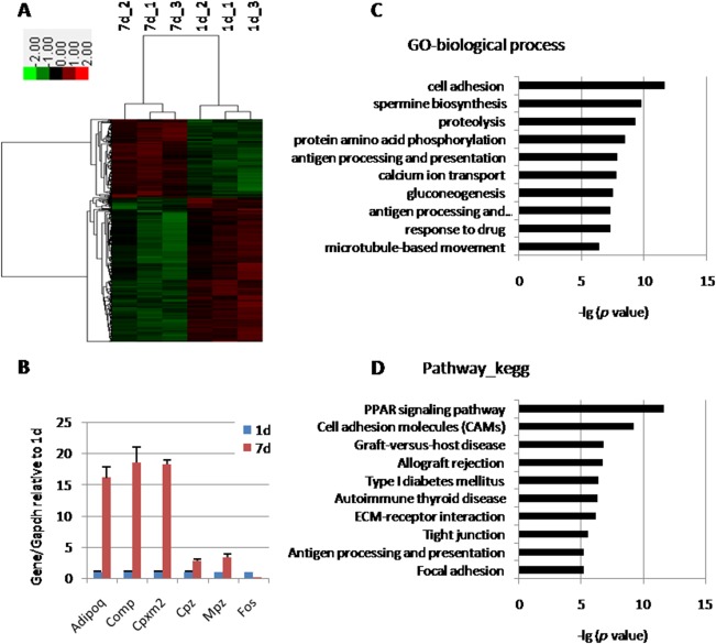
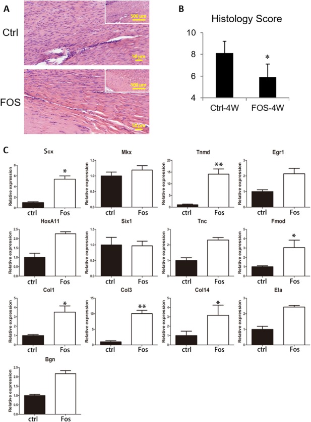
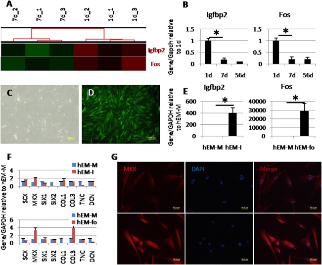
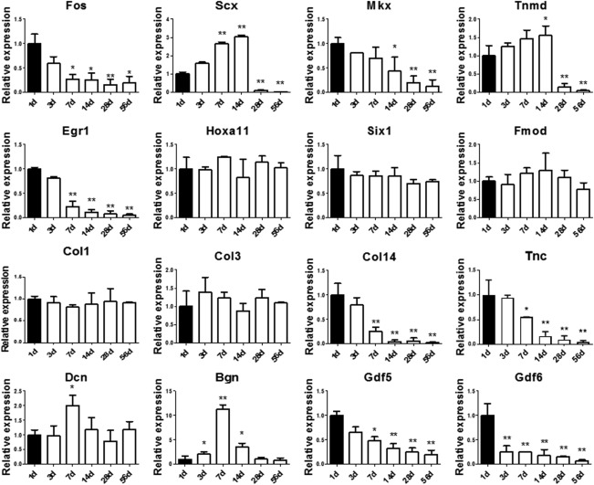
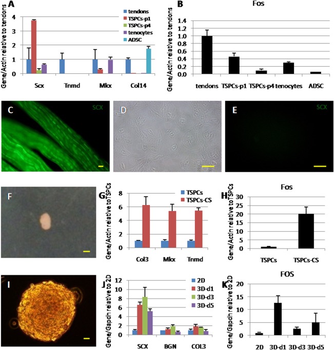
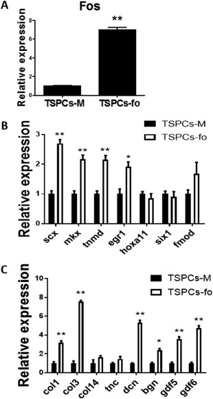
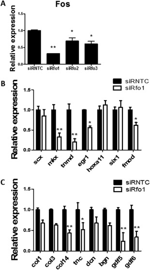
Stem cells have been widely used in tendon tissue engineering. The lack of refined and controlled differentiation strategy hampers the tendon repair and regeneration. This study aimed to find new effective differentiation factors for stepwise tenogenic differentiation. By microarray screening, the transcript factor Fos was found to be expressed in significantly higher amounts in postnatal Achilles tendon tissue derived from 1 day as compared with 7-days-old rats. It was further confirmed that expression of Fos decreased with time in postnatal rat Achilles tendon, which was accompanied with the decreased expression of multiply tendon markers. The expression of Fos also declined during regular in vitro cell culture, which corresponded to the loss of tendon phenotype. In a cell-sheet and a three-dimensional cell culture model, the expression of Fos was upregulated as compared with in regular cell culture, together with the recovery of tendon phenotype. In addition, significant higher expression of tendon markers was found in Fos-overexpressed tendon stem/progenitor cells (TSPCs), and Fos knock-down gave opposite results. In situ rat tendon repair experiments found more normal tendon-like tissue formed and higher tendon markers expression at 4 weeks postimplantation of Fos-overexpressed TSPCs derived nonscaffold engineering tendon (cell-sheet), as compared with the control group. This study identifies Fos as a new marker and functional driver in the early stage teno-lineage differentiation of tendon, which paves the way for effective stepwise tendon differentiation and future tendon regeneration. Stem Cells Translational Medicine 2017;6:2009-2019.
干细胞在肌腱组织工程中得到了广泛应用。缺乏精细和可控的分化策略阻碍了肌腱的修复和再生。本研究旨在寻找新的有效的分化因子,以逐步进行肌腱向分化。通过微阵列筛选,发现转录因子 Fos 在 1 天龄大鼠的比目鱼肌腱组织中表达量明显高于 7 天龄大鼠。进一步证实 Fos 的表达在出生后大鼠跟腱中随时间的推移而降低,同时伴有多重肌腱标记物的表达降低。Fos 的表达也在常规细胞培养过程中下降,这与肌腱表型的丧失相对应。在细胞片层和三维细胞培养模型中,与常规细胞培养相比,Fos 的表达上调,同时恢复了肌腱表型。此外,在 Fos 过表达的肌腱干/祖细胞 (TSPCs) 中发现了更高水平的肌腱标记物表达,而 Fos 敲低则得到了相反的结果。在原位大鼠肌腱修复实验中发现,与对照组相比,Fos 过表达的 TSPCs 衍生的无支架工程肌腱 (细胞片层) 植入后 4 周形成了更多正常的肌腱样组织,且肌腱标记物表达更高。本研究鉴定出 Fos 是肌腱早期向肌腱谱系分化的新标记物和功能驱动因子,为有效的逐步肌腱分化和未来的肌腱再生铺平了道路。干细胞转化医学 2017;6:2009-2019。