Terry James G, Shay Christina M, Schreiner Pamela J, Jacobs David R, Sanchez Otto A, Reis Jared P, Goff David C, Gidding Samuel S, Steffen Lyn M, Carr John Jeffrey
From the Department of Radiology, Vanderbilt University School of Medicine, Nashville, TN (J.G.T., J.J.C.); American Heart Association, Dallas, TX (C.M.S.); School of Public Health (P.J.S., D.R.J., L.M.S.) and Department of Medicine (O.A.S.), University of Minnesota, Minneapolis; Department of Internal Medicine (O.A.S.); National Institutes of Health, Bethesda, MD (J.P.R., D.C.G.); and A.I. DuPont Hospital of Children, Wilmington, DE (S.S.G.).
Arterioscler Thromb Vasc Biol. 2017 Dec;37(12):2370-2378. doi: 10.1161/ATVBAHA.117.309633. Epub 2017 Oct 12.
Excess deposition of fat within and around vital organs and nonadipose tissues is hypothesized to contribute to cardiovascular disease (CVD) risk. We evaluated the association of abdominal intermuscular adipose tissue (IMAT) volume with coronary artery calcification in the CARDIA study (Coronary Artery Risk Development in Young Adults) participants.
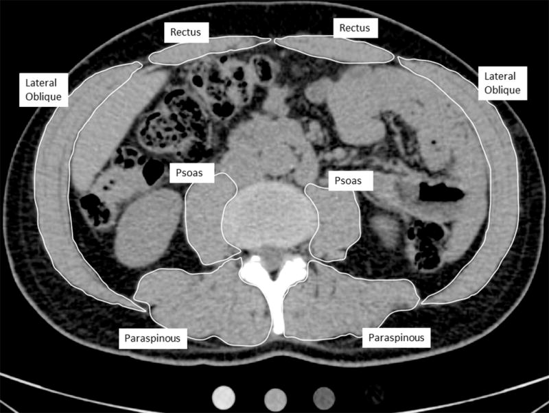
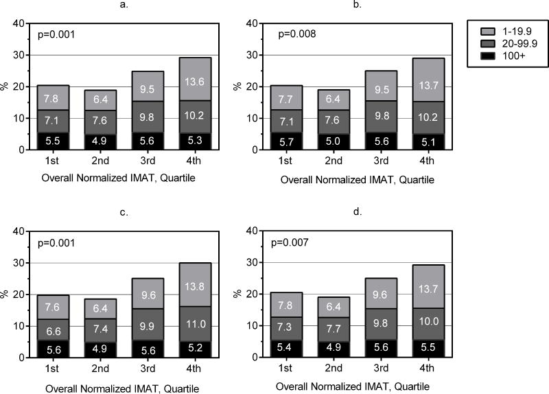
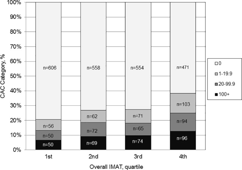
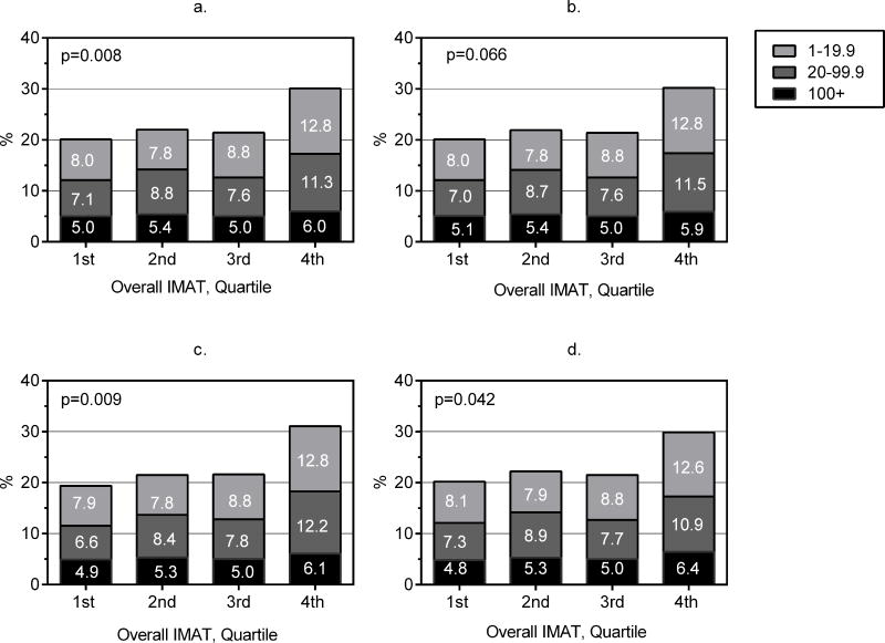
We measured IMAT in the abdominal muscles, visceral adipose tissue and pericardial adipose tissue, and coronary artery calcification using computed tomography in 3051 CARDIA participants (56% women) at the CARDIA year 25 examination (2010-2011). Mean IMAT volume and mean IMAT/total muscle volume (IMAT normalized for muscle size) were calculated in a 10-mm block of slices centered at L3-L4. Multivariable analyses included potential confounders and traditional cardiovascular disease risk factors. Compared with the lowest quartile, the upper quartile of abdominal IMAT volume was associated with higher coronary artery calcification prevalence (odds ratio [95% confidence interval], 1.6 [1.2-2.1]) after adjusting for cardiovascular disease risk factors. Results were similar for highest versus lowest quartile of IMAT normalized to total muscle volume (odds ratio [95% confidence interval], 1.5 [1.1-2.0]). Significant associations of higher IMAT and normalized IMAT with coronary artery calcification prevalence persisted when body mass index, visceral adipose tissue, or pericardial adipose tissue were added to the models.
In a large, community-based, cross-sectional study, we found that higher abdominal skeletal muscle adipose tissue volume was associated with subclinical atherosclerosis independent of traditional cardiovascular disease risk factors and other adipose depots.
据推测,重要器官内部及周围以及非脂肪组织中脂肪过度沉积会增加心血管疾病(CVD)风险。在青年动脉粥样硬化风险发展研究(CARDIA研究)参与者中,我们评估了腹部肌间脂肪组织(IMAT)体积与冠状动脉钙化之间的关联。
在CARDIA研究第25年(2010 - 2011年)检查时,我们对3051名CARDIA参与者(56%为女性)使用计算机断层扫描测量了腹部肌肉中的IMAT、内脏脂肪组织和心包脂肪组织以及冠状动脉钙化情况。在以L3 - L4为中心的10毫米切片块中计算平均IMAT体积和平均IMAT/总肌肉体积(根据肌肉大小进行标准化的IMAT)。多变量分析纳入了潜在混杂因素和传统心血管疾病风险因素。在调整心血管疾病风险因素后,与最低四分位数相比,腹部IMAT体积的最高四分位数与更高的冠状动脉钙化患病率相关(优势比[95%置信区间],1.6[1.2 - 2.1])。对于标准化至总肌肉体积的IMAT的最高与最低四分位数,结果相似(优势比[95%置信区间],1.5[1.1 - 2.0])。当将体重指数、内脏脂肪组织或心包脂肪组织添加到模型中时,较高的IMAT和标准化IMAT与冠状动脉钙化患病率之间的显著关联仍然存在。
在一项基于社区的大型横断面研究中,我们发现较高的腹部骨骼肌脂肪组织体积与亚临床动脉粥样硬化相关,且独立于传统心血管疾病风险因素和其他脂肪储存部位。