• 文献检索
  • 文档翻译
  • 深度研究
  • 学术资讯
  • Suppr Zotero 插件Zotero 插件
  • 邀请有礼
  • 套餐&价格
  • 历史记录
应用&插件
Suppr Zotero 插件Zotero 插件浏览器插件Mac 客户端Windows 客户端微信小程序
定价
高级版会员购买积分包购买API积分包
服务
文献检索文档翻译深度研究API 文档MCP 服务
关于我们
关于 Suppr公司介绍联系我们用户协议隐私条款
关注我们

Suppr 超能文献

核心技术专利:CN118964589B侵权必究
粤ICP备2023148730 号-1Suppr @ 2026

文献检索

告别复杂PubMed语法,用中文像聊天一样搜索,搜遍4000万医学文献。AI智能推荐,让科研检索更轻松。

立即免费搜索

文件翻译

保留排版,准确专业,支持PDF/Word/PPT等文件格式,支持 12+语言互译。

免费翻译文档

深度研究

AI帮你快速写综述,25分钟生成高质量综述,智能提取关键信息,辅助科研写作。

立即免费体验

糖尿病视网膜病变患者全视网膜光凝术后视盘周围神经组织的定量分析。

Quantitative analysis of neural tissues around the optic disc after panretinal photocoagulation in patients with diabetic retinopathy.

作者信息

Yang Hyun Seung, Kim June-Gone, Cha Jae Bong, Yun Young In, Park Jong Hoon, Woo Jong Eun

机构信息

Department of Ophthalmology, Seoul Shinsegae Eye Center, Eui Jung Bu, Gyeonggi-do, South Korea.

Department of Ophthalmology, University of Ulsan, College of Medicine, Asan Medical Center, Seoul, South Korea.

出版信息

PLoS One. 2017 Oct 17;12(10):e0186229. doi: 10.1371/journal.pone.0186229. eCollection 2017.

DOI:10.1371/journal.pone.0186229
PMID:29040280
原文链接:https://pmc.ncbi.nlm.nih.gov/articles/PMC5645105/
Abstract

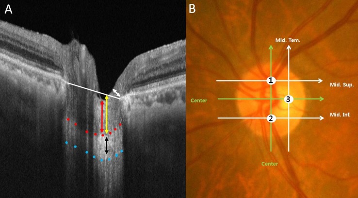
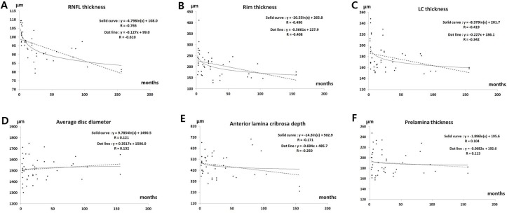
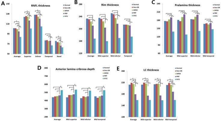
In this retrospective cross-sectional study, we quantitatively analyzed the tomographic features in the neural tissues around the optic disc in patients with diabetic retinopathy with and without panretinal photocoagulation. We analyzed 206 eyes, comprising 33 normal eyes in subjects without diabetes (group I), 30 eyes without diabetic retinopathy (group II), 66 eyes with non-proliferative diabetic retinopathy (group III), 45 eyes with panretinal photocoagulation (group IV), and 32 eyes with normal tension glaucoma (group V). Sequential images acquired using swept-source optical coherence tomography in three-dimensional mode were used to measure peripapillary retinal nerve fiber layer thickness, neuro-retinal rim thickness, anterior lamina cribrosa depth, prelaminar thickness, and thickness of the lamina cribrosa. The peripapillary retinal nerve fiber layer thickness and lamina cribrosa thickness were significantly thinner in group IV than in group III (p = 0.019 and p < 0.001). However, there was no significant difference in rim thickness, anterior lamina cribrosa depth, or prelaminar thickness between groups III and IV (p = 0.307, p = 0.877, and p = 0.212). Multivariate analysis revealed that time since panretinal photocoagulation and thickness of the lamina cribrosa had a significant effect on peripapillary retinal nerve fiber layer thickness (p < 0.001 and p = 0.014). In group IV, there was a negative correlation between time elapsed since panretinal photocoagulation and peripapillary retinal nerve fiber layer thickness, rim thickness, and thickness of the lamina cribrosa (r = -0.765, r = -0.490, and r = -0.419), but no correlation with prelaminar thickness or anterior lamina cribrosa depth (r = 0.104 and r = -0.171). Panretinal photocoagulation may be related to thinning of the peripapillary retinal nerve fiber layer, rim thickness, and lamina cribrosa, but not prelaminar thickness or anterior lamina cribrosa depth. These features are different from the peripapillary features of eyes with typical normal tension glaucoma.

摘要

在这项回顾性横断面研究中,我们定量分析了接受全视网膜光凝和未接受全视网膜光凝的糖尿病视网膜病变患者视盘周围神经组织的断层特征。我们分析了206只眼睛,包括无糖尿病受试者的33只正常眼睛(I组)、30只无糖尿病视网膜病变的眼睛(II组)、66只非增殖性糖尿病视网膜病变的眼睛(III组)、45只接受全视网膜光凝的眼睛(IV组)以及32只正常眼压性青光眼的眼睛(V组)。使用扫频源光学相干断层扫描以三维模式获取的序列图像用于测量视乳头周围视网膜神经纤维层厚度、神经视网膜边缘厚度、筛板前深度、板前厚度和筛板厚度。IV组的视乳头周围视网膜神经纤维层厚度和筛板厚度明显薄于III组(p = 0.019和p < 0.001)。然而,III组和IV组之间的边缘厚度、筛板前深度或板前厚度没有显著差异(p = 0.307、p = 0.877和p = 0.212)。多变量分析显示,全视网膜光凝后的时间和筛板厚度对视乳头周围视网膜神经纤维层厚度有显著影响(p < 0.001和p = 0.014)。在IV组中,全视网膜光凝后的时间与视乳头周围视网膜神经纤维层厚度、边缘厚度和筛板厚度呈负相关(r = -0.765、r = -0.490和r = -0.419),但与板前厚度或筛板前深度无相关性(r = 0.104和r = -0.171)。全视网膜光凝可能与视乳头周围视网膜神经纤维层、边缘厚度和筛板变薄有关,但与板前厚度或筛板前深度无关。这些特征与典型正常眼压性青光眼眼睛的视乳头周围特征不同。

https://cdn.ncbi.nlm.nih.gov/pmc/blobs/1a6a/5645105/55aeda13ccb8/pone.0186229.g003.jpg
https://cdn.ncbi.nlm.nih.gov/pmc/blobs/1a6a/5645105/e4b1337bafc5/pone.0186229.g001.jpg
https://cdn.ncbi.nlm.nih.gov/pmc/blobs/1a6a/5645105/ebb6a09983c1/pone.0186229.g002.jpg
https://cdn.ncbi.nlm.nih.gov/pmc/blobs/1a6a/5645105/55aeda13ccb8/pone.0186229.g003.jpg
https://cdn.ncbi.nlm.nih.gov/pmc/blobs/1a6a/5645105/e4b1337bafc5/pone.0186229.g001.jpg
https://cdn.ncbi.nlm.nih.gov/pmc/blobs/1a6a/5645105/ebb6a09983c1/pone.0186229.g002.jpg
https://cdn.ncbi.nlm.nih.gov/pmc/blobs/1a6a/5645105/55aeda13ccb8/pone.0186229.g003.jpg

相似文献

1
Quantitative analysis of neural tissues around the optic disc after panretinal photocoagulation in patients with diabetic retinopathy.糖尿病视网膜病变患者全视网膜光凝术后视盘周围神经组织的定量分析。
PLoS One. 2017 Oct 17;12(10):e0186229. doi: 10.1371/journal.pone.0186229. eCollection 2017.
2
Circumpapillary retinal nerve fiber layer thickness, anterior lamina cribrosa depth, and lamina cribrosa thickness in neovascular glaucoma secondary to proliferative diabetic retinopathy: a cross-sectional study.增殖性糖尿病视网膜病变继发新生血管性青光眼中的视乳头周围视网膜神经纤维层厚度、筛板前深度和筛板厚度:一项横断面研究
BMC Ophthalmol. 2017 Apr 26;17(1):57. doi: 10.1186/s12886-017-0456-9.
3
Lamina cribrosa position and Bruch's membrane opening differences between anterior ischemic optic neuropathy and open-angle glaucoma.前部缺血性视神经病变与开角型青光眼之间的筛板位置及布鲁赫膜开口差异
Eur J Ophthalmol. 2019 Mar;29(2):202-209. doi: 10.1177/1120672118782101. Epub 2018 Jun 18.
4
Evaluation of the lamina cribrosa in patients with diabetes mellitus using enhanced depth imaging spectral-domain optical coherence tomography.使用增强深度成像光谱域光学相干断层扫描技术评估糖尿病患者的筛板。
Diab Vasc Dis Res. 2018 Sep;15(5):442-448. doi: 10.1177/1479164118782091. Epub 2018 Jun 12.
5
Long-term Effect of Panretinal Photocoagulation on Spectral Domain Optical Coherence Tomography Measurements in Diabetic Retinopathy.全视网膜光凝对糖尿病视网膜病变光谱域光学相干断层扫描测量的长期影响
Curr Eye Res. 2017 Aug;42(8):1169-1173. doi: 10.1080/02713683.2017.1280510. Epub 2017 Mar 24.
6
PANRETINAL PHOTOCOAGULATION VERSUS RANIBIZUMAB FOR PROLIFERATIVE DIABETIC RETINOPATHY: Comparison of Peripapillary Retinal Nerve Fiber Layer Thickness in a Randomized Clinical Trial.全视网膜光凝与雷珠单抗治疗增生性糖尿病视网膜病变的比较:一项随机临床试验中视盘周围视网膜神经纤维层厚度的比较。
Retina. 2019 Jan;39(1):69-78. doi: 10.1097/IAE.0000000000001909.
7
Changes in Macular Retinal Layers and Peripapillary Nerve Fiber Layer Thickness after 577-nm Pattern Scanning Laser in Patients with Diabetic Retinopathy.糖尿病视网膜病变患者接受577纳米模式扫描激光治疗后黄斑视网膜各层及视乳头周围神经纤维层厚度的变化
Korean J Ophthalmol. 2017 Dec;31(6):497-507. doi: 10.3341/kjo.2016.0108. Epub 2017 Nov 30.
8
Posterior displacement of the lamina cribrosa in normal-tension and high-tension glaucoma.正常眼压性青光眼和高眼压性青光眼患者筛板的后移位
Acta Ophthalmol. 2016 Sep;94(6):e492-500. doi: 10.1111/aos.13012. Epub 2016 Mar 24.
9
Randomized clinical trial to evaluate the effects of Pascal panretinal photocoagulation on macular nerve fiber layer: Manchester Pascal Study report 3.随机临床试验评估帕斯卡全视网膜光凝对黄斑神经纤维层的影响:曼彻斯特帕斯卡研究报告 3。
Retina. 2011 Sep;31(8):1699-707. doi: 10.1097/IAE.0b013e318207d188.
10
Optic nerve head cupping in glaucomatous and non-glaucomatous optic neuropathy.青光眼性和非青光眼性视神经病变的视神经头凹陷。
Br J Ophthalmol. 2019 Mar;103(3):374-378. doi: 10.1136/bjophthalmol-2018-312161. Epub 2018 May 23.

引用本文的文献

1
Effect of panretinal photocoagulation versus intravitreal bevacizumab injection on optic disc microcirculation in patients with diabetic retinopathy.全视网膜光凝与玻璃体腔注射贝伐单抗对糖尿病视网膜病变患者视盘微循环的影响。
Int J Retina Vitreous. 2024 Dec 18;10(1):98. doi: 10.1186/s40942-024-00621-w.
2
Did you see it? A Python tool for psychophysical assessment of the human blind spot.你看到了吗?一个用于人类盲点心理物理评估的 Python 工具。
PLoS One. 2021 Nov 4;16(11):e0254195. doi: 10.1371/journal.pone.0254195. eCollection 2021.
3
Optical Coherence Tomography Imaging of the Lamina Cribrosa: Structural Biomarkers in Nonglaucomatous Diseases.

本文引用的文献

1
Influence of Large Intraocular Pressure Reduction on Peripapillary OCT Vessel Density in Ocular Hypertensive and Glaucoma Eyes.眼压大幅降低对高眼压症和青光眼患者视乳头周围光学相干断层扫描血管密度的影响。
J Glaucoma. 2017 Jan;26(1):e7-e10. doi: 10.1097/IJG.0000000000000527.
2
Vessel density calculated from OCT angiography in 3 peripapillary sectors in normal, ocular hypertensive, and glaucoma eyes.通过光学相干断层扫描血管造影术计算正常眼、高眼压症眼和青光眼眼中3个视乳头周围区域的血管密度。
Eur J Ophthalmol. 2016 Apr 12;26(3):e42-5. doi: 10.5301/ejo.5000717.
3
Influence of translaminar pressure dynamics on the position of the anterior lamina cribrosa surface.
筛板的光学相干断层扫描成像:非青光眼性疾病中的结构生物标志物
J Ophthalmol. 2021 Feb 19;2021:8844614. doi: 10.1155/2021/8844614. eCollection 2021.
4
Long-term effects of pattern scan laser pan-retinal photocoagulation on diabetic retinopathy in Chinese patients: a retrospective study.模式扫描激光全视网膜光凝术对中国患者糖尿病视网膜病变的长期影响:一项回顾性研究
Int J Ophthalmol. 2020 Feb 18;13(2):239-245. doi: 10.18240/ijo.2020.02.06. eCollection 2020.
5
Down-regulation of miR-377 suppresses high glucose and hypoxia-induced angiogenesis and inflammation in human retinal endothelial cells by direct up-regulation of target gene SIRT1.下调 miR-377 通过直接上调靶基因 SIRT1 抑制高糖和低氧诱导的人视网膜内皮细胞血管生成和炎症反应。
Hum Cell. 2019 Jul;32(3):260-274. doi: 10.1007/s13577-019-00240-w. Epub 2019 Jan 31.
跨筛板压力动态变化对筛板前表面位置的影响。
Invest Ophthalmol Vis Sci. 2015 May;56(5):2833-41. doi: 10.1167/iovs.14-15869.
4
Anterior lamina cribrosa insertion in primary open-angle glaucoma patients and healthy subjects.原发性开角型青光眼患者和健康受试者的筛板前插入部
PLoS One. 2014 Dec 22;9(12):e114935. doi: 10.1371/journal.pone.0114935. eCollection 2014.
5
Comparison of lamina cribrosa thickness in normal tension glaucoma patients with unilateral visual field defect.正常眼压性青光眼单侧视野缺损患者筛板厚度的比较。
Am J Ophthalmol. 2015 Mar;159(3):512-8.e1. doi: 10.1016/j.ajo.2014.11.034. Epub 2014 Dec 9.
6
Comparison of the thickness of the lamina cribrosa and vascular factors in early normal-tension glaucoma with low and high intraocular pressures.早期正常眼压性青光眼伴低眼压和高眼压时筛板厚度与血管因素的比较。
Korean J Ophthalmol. 2014 Dec;28(6):473-8. doi: 10.3341/kjo.2014.28.6.473. Epub 2014 Nov 19.
7
Influence of lamina cribrosa thickness and depth on the rate of progressive retinal nerve fiber layer thinning.视盘筛板厚度和深度对视网膜神经纤维层进行性变薄速率的影响。
Ophthalmology. 2015 Apr;122(4):721-9. doi: 10.1016/j.ophtha.2014.10.007. Epub 2014 Nov 26.
8
Changes in peripapillary retinal nerve fiber layer thickness after pattern scanning laser photocoagulation in patients with diabetic retinopathy.糖尿病性视网膜病变患者经模式扫描激光光凝治疗后视乳头周围视网膜神经纤维层厚度的变化
Korean J Ophthalmol. 2014 Jun;28(3):220-5. doi: 10.3341/kjo.2014.28.3.220. Epub 2014 May 19.
9
Effect of diabetic macular edema on peripapillary retinal nerve fiber layer thickness profiles.糖尿病性黄斑水肿对视盘周围视网膜神经纤维层厚度曲线的影响。
Invest Ophthalmol Vis Sci. 2014 May 15;55(7):4213-9. doi: 10.1167/iovs.13-13776.
10
Change of retinal nerve fiber layer thickness in various retinal diseases treated with multiple intravitreal antivascular endothelial growth factor.多种抗血管内皮生长因子眼内注射治疗各种视网膜疾病后视网膜神经纤维层厚度的变化。
Invest Ophthalmol Vis Sci. 2014 Apr 15;55(4):2403-11. doi: 10.1167/iovs.13-13769.