• 文献检索
  • 文档翻译
  • 深度研究
  • 学术资讯
  • Suppr Zotero 插件Zotero 插件
  • 邀请有礼
  • 套餐&价格
  • 历史记录
应用&插件
Suppr Zotero 插件Zotero 插件浏览器插件Mac 客户端Windows 客户端微信小程序
定价
高级版会员购买积分包购买API积分包
服务
文献检索文档翻译深度研究API 文档MCP 服务
关于我们
关于 Suppr公司介绍联系我们用户协议隐私条款
关注我们

Suppr 超能文献

核心技术专利:CN118964589B侵权必究
粤ICP备2023148730 号-1Suppr @ 2026

文献检索

告别复杂PubMed语法,用中文像聊天一样搜索,搜遍4000万医学文献。AI智能推荐,让科研检索更轻松。

立即免费搜索

文件翻译

保留排版,准确专业,支持PDF/Word/PPT等文件格式,支持 12+语言互译。

免费翻译文档

深度研究

AI帮你快速写综述,25分钟生成高质量综述,智能提取关键信息,辅助科研写作。

立即免费体验

患有冲动性攻击的参战退役军人的静息态功能连接。

Resting-state functional connectivity in combat veterans suffering from impulsive aggression.

机构信息

University Medical Center, Heidelberglaan 100, 3584 CX Utrecht, The Netherlands.

Research Center Military Mental Health Care, P.O. Box 90000, 3509 AA, Utrecht, The Netherlands.

出版信息

Soc Cogn Affect Neurosci. 2017 Dec 1;12(12):1881-1889. doi: 10.1093/scan/nsx113.

DOI:10.1093/scan/nsx113
PMID:29040723
原文链接:https://pmc.ncbi.nlm.nih.gov/articles/PMC5716169/
Abstract

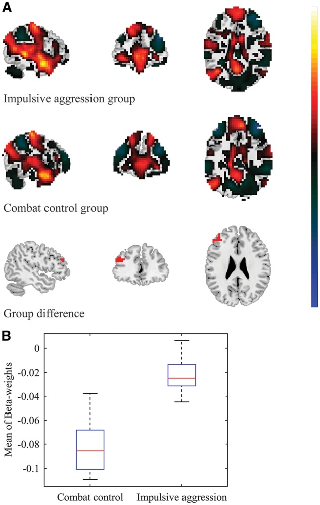
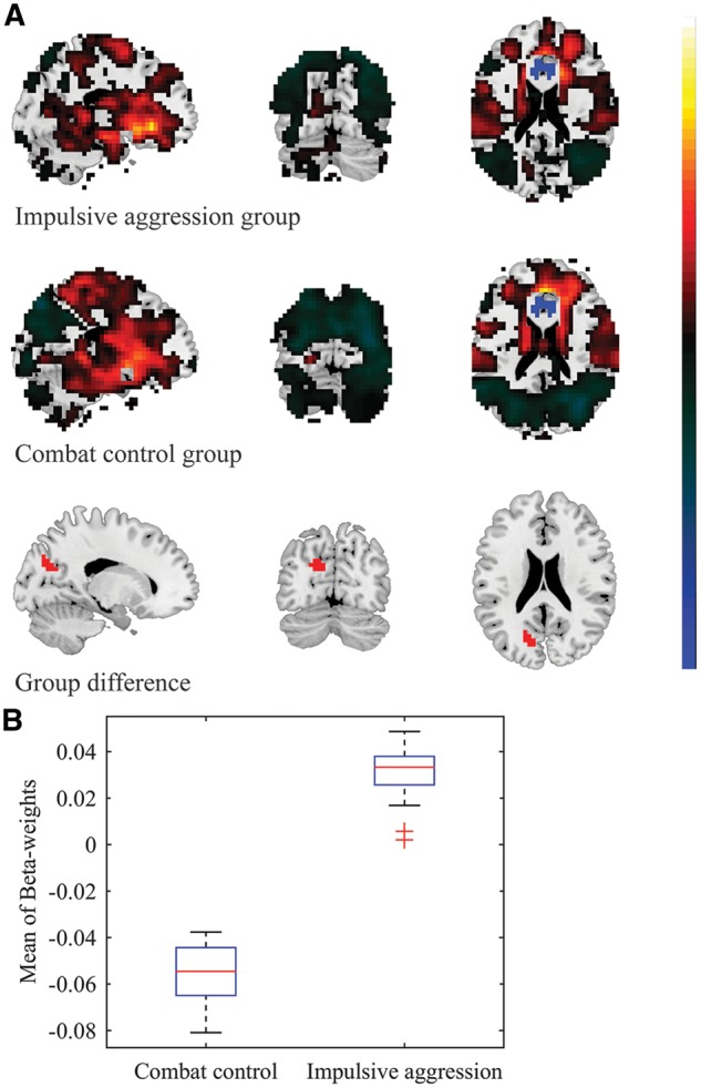
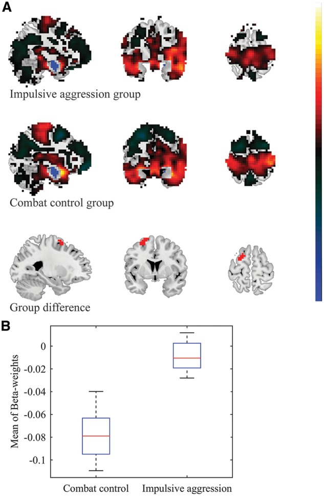
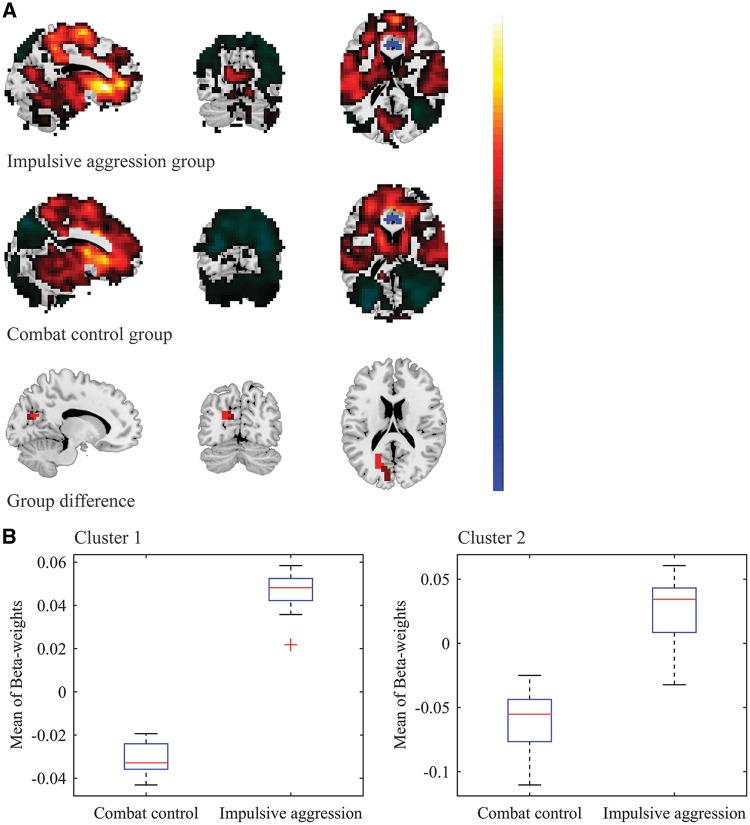
Impulsive aggression is common among military personnel after deployment and may arise because of impaired top-down regulation of the amygdala by prefrontal regions. This study sought to further explore this hypothesis via resting-state functional connectivity analyses in impulsively aggressive combat veterans. Male combat veterans with (n = 28) and without (n = 30) impulsive aggression problems underwent resting-state functional magnetic resonance imaging. Functional connectivity analyses were conducted with the following seed-regions: basolateral amygdala (BLA), centromedial amygdala, anterior cingulate cortex (ACC), and anterior insular cortex (AIC). Regions-of-interest analyses focused on the orbitofrontal cortex and periaqueductal gray, and yielded no significant results. In exploratory cluster analyses, we observed reduced functional connectivity between the (bilateral) BLA and left dorsolateral prefrontal cortex in the impulsive aggression group, relative to combat controls. This finding indicates that combat-related impulsive aggression may be marked by weakened functional connectivity between the amygdala and prefrontal regions, already in the absence of explicit emotional stimuli. Group differences in functional connectivity were also observed between the (bilateral) ACC and left cuneus, which may be related to heightened vigilance to potentially threatening visual cues, as well as between the left AIC and right temporal pole, possibly related to negative memory association in impulsive aggression.

摘要

冲动性攻击在部署后的军事人员中很常见,可能是由于前额叶区域对杏仁核的自上而下调节受损所致。本研究通过对冲动性攻击的战斗退伍军人进行静息态功能连接分析,进一步探讨了这一假设。有(n=28)和没有(n=30)冲动性攻击问题的男性战斗退伍军人接受了静息态功能磁共振成像。使用以下种子区域进行功能连接分析:基底外侧杏仁核(BLA)、中央杏仁核、前扣带皮层(ACC)和前岛叶皮层(AIC)。重点关注眶额皮层和导水管周围灰质的 ROI 分析,未得到显著结果。在探索性聚类分析中,我们观察到冲动攻击组中(双侧)BLA 与左背外侧前额叶皮层之间的功能连接降低,与战斗对照组相比。这一发现表明,与战斗相关的冲动性攻击可能以杏仁核与前额叶区域之间的功能连接减弱为特征,即使在没有明确的情绪刺激的情况下也是如此。冲动性攻击组和对照组之间在(双侧)ACC 和左侧楔前叶之间也观察到功能连接的差异,这可能与对潜在威胁性视觉线索的警惕性增加有关,而在左侧 AIC 和右侧颞极之间也可能与冲动性攻击中的负性记忆联想有关。

https://cdn.ncbi.nlm.nih.gov/pmc/blobs/f878/5716169/105077b1829f/nsx113f4.jpg
https://cdn.ncbi.nlm.nih.gov/pmc/blobs/f878/5716169/9ff691d4a393/nsx113f1.jpg
https://cdn.ncbi.nlm.nih.gov/pmc/blobs/f878/5716169/decde1d01592/nsx113f2.jpg
https://cdn.ncbi.nlm.nih.gov/pmc/blobs/f878/5716169/e231fa596609/nsx113f3.jpg
https://cdn.ncbi.nlm.nih.gov/pmc/blobs/f878/5716169/105077b1829f/nsx113f4.jpg

相似文献

1
Resting-state functional connectivity in combat veterans suffering from impulsive aggression.患有冲动性攻击的参战退役军人的静息态功能连接。
Soc Cogn Affect Neurosci. 2017 Dec 1;12(12):1881-1889. doi: 10.1093/scan/nsx113.
2
Voxel-based morphometry and cortical thickness in combat veterans suffering from impulsive aggression.基于体素的形态测量学和冲动性攻击的参战退役军人的皮质厚度。
Psychol Med. 2021 Jun;51(8):1299-1309. doi: 10.1017/S0033291720000033. Epub 2020 Feb 7.
3
Resting state functional connectivity of the anterior cingulate cortex in veterans with and without post-traumatic stress disorder.患有和未患有创伤后应激障碍的退伍军人前扣带回皮质的静息态功能连接性
Hum Brain Mapp. 2015 Jan;36(1):99-109. doi: 10.1002/hbm.22615. Epub 2014 Aug 19.
4
Real-time fMRI amygdala neurofeedback positive emotional training normalized resting-state functional connectivity in combat veterans with and without PTSD: a connectome-wide investigation.实时 fMRI 杏仁核神经反馈正性情绪训练对 PTSD 及非 PTSD 参战退役军人静息态功能连接的归一化作用:一项连接组学研究。
Neuroimage Clin. 2018 Aug 19;20:543-555. doi: 10.1016/j.nicl.2018.08.025. eCollection 2018.
5
Sex differences in orbitofrontal connectivity in male and female veterans with TBI.患有创伤性脑损伤的男性和女性退伍军人眶额连接的性别差异。
Brain Imaging Behav. 2015 Sep;9(3):535-49. doi: 10.1007/s11682-015-9379-3.
6
Altered resting-state functional connectivity of basolateral and centromedial amygdala complexes in posttraumatic stress disorder.创伤后应激障碍患者基底外侧和中央杏仁核复合体的静息态功能连接改变。
Neuropsychopharmacology. 2014 Jan;39(2):351-9. doi: 10.1038/npp.2013.197. Epub 2013 Aug 9.
7
ALTERED DEFAULT MODE NETWORK (DMN) RESTING STATE FUNCTIONAL CONNECTIVITY FOLLOWING A MINDFULNESS-BASED EXPOSURE THERAPY FOR POSTTRAUMATIC STRESS DISORDER (PTSD) IN COMBAT VETERANS OF AFGHANISTAN AND IRAQ.基于正念的暴露疗法对阿富汗和伊拉克战争退伍军人创伤后应激障碍(PTSD)的影响:默认模式网络(DMN)静息态功能连接的改变
Depress Anxiety. 2016 Apr;33(4):289-99. doi: 10.1002/da.22481.
8
Anger provocation increases limbic and decreases medial prefrontal cortex connectivity with the left amygdala in reactive aggressive violent offenders.愤怒激发会增加反应性攻击暴力罪犯的边缘系统与左侧杏仁核的连接,同时减少内侧前额叶皮质的连接。
Brain Imaging Behav. 2019 Oct;13(5):1311-1323. doi: 10.1007/s11682-018-9945-6.
9
Altered resting-state amygdala functional connectivity in men with posttraumatic stress disorder.创伤后应激障碍男性静息状态杏仁核功能连接改变。
J Psychiatry Neurosci. 2012 Jul;37(4):241-9. doi: 10.1503/jpn.110069.
10
Neural activity during the viewing of emotional pictures in veterans with pathological anger and aggression.病理性愤怒和攻击的退伍军人观看情绪图片时的神经活动。
Eur Psychiatry. 2018 Jan;47:1-8. doi: 10.1016/j.eurpsy.2017.09.002. Epub 2017 Sep 22.

引用本文的文献

1
Functional cerebello-cortico-limbic connectivity in aggression: A resting-state 7T fMRI study in healthy volunteers.攻击行为中的功能性小脑-皮质-边缘系统连接:一项针对健康志愿者的静息态7T功能磁共振成像研究。
Imaging Neurosci (Camb). 2025 Jun 20;3. doi: 10.1162/IMAG.a.42. eCollection 2025.
2
Deciphering the Neural Effects of Emotional, Motivational, and Cognitive Challenges on Inhibitory Control Processes.解读情绪、动机和认知挑战对抑制控制过程的神经影响。
Hum Brain Mapp. 2025 Feb 1;46(2):e70137. doi: 10.1002/hbm.70137.
3
Amygdala fMRI-A Critical Appraisal of the Extant Literature.

本文引用的文献

1
Disrupted functional connectivity in adolescent obesity.青少年肥胖中功能连接的破坏。
Neuroimage Clin. 2016 Jul 12;12:262-8. doi: 10.1016/j.nicl.2016.07.005. eCollection 2016.
2
A landscape-based cluster analysis using recursive search instead of a threshold parameter.一种基于景观的聚类分析,使用递归搜索而非阈值参数。
MethodsX. 2016 Jul 7;3:477-82. doi: 10.1016/j.mex.2016.06.002. eCollection 2016.
3
Amygdala hyperactivation to angry faces in intermittent explosive disorder.间歇性暴发性障碍患者杏仁核对于愤怒面孔的过度激活。
杏仁核功能磁共振成像——对现有文献的批判性评价
Neurosci Insights. 2024 Aug 13;19:26331055241270591. doi: 10.1177/26331055241270591. eCollection 2024.
4
The role of intimate partner violence perpetrators' resting state functional connectivity in treatment compliance and recidivism.亲密伴侣暴力行为者静息态功能连接在治疗依从性和复发性中的作用。
Sci Rep. 2024 Jan 30;14(1):2472. doi: 10.1038/s41598-024-52443-3.
5
Pattern classification based on the amygdala does not predict an individual's response to emotional stimuli.基于杏仁核的模式分类不能预测个体对情绪刺激的反应。
Hum Brain Mapp. 2023 Aug 15;44(12):4452-4466. doi: 10.1002/hbm.26391. Epub 2023 Jun 23.
6
Neurobiology of Aggression-Review of Recent Findings and Relationship with Alcohol and Trauma.攻击行为的神经生物学——近期研究发现及其与酒精和创伤的关系综述
Biology (Basel). 2023 Mar 20;12(3):469. doi: 10.3390/biology12030469.
7
Trait Aggression is Reflected by a Lower Temporal Stability of EEG Resting Networks.特质攻击性表现为 EEG 静息网络的时间稳定性降低。
Brain Topogr. 2024 Jul;37(4):514-523. doi: 10.1007/s10548-022-00929-6. Epub 2022 Nov 18.
8
Increased amygdala and decreased frontolimbic r esting- s tate functional connectivity in children with aggressive behavior.具有攻击性行为儿童的杏仁核和额眶额回静息态功能连接增加。
Soc Cogn Affect Neurosci. 2022 Jul 2;17(7):634-644. doi: 10.1093/scan/nsab128.
9
Functional connectivity of amygdala subregions predicts vulnerability to depression following the COVID-19 pandemic.杏仁核亚区的功能连接可预测 COVID-19 大流行后个体对抑郁的易感性。
J Affect Disord. 2022 Jan 15;297:421-429. doi: 10.1016/j.jad.2021.09.107. Epub 2021 Oct 2.
10
Neural correlates of anger expression in patients with PTSD.创伤后应激障碍患者愤怒表达的神经相关性。
Neuropsychopharmacology. 2021 Aug;46(9):1635-1642. doi: 10.1038/s41386-020-00942-y. Epub 2021 Jan 26.
J Psychiatr Res. 2016 Aug;79:34-41. doi: 10.1016/j.jpsychires.2016.04.006. Epub 2016 Apr 24.
4
Cytoarchitecture and probability maps of the human medial orbitofrontal cortex.人类内侧眶额皮质的细胞构筑和概率图谱。
Cortex. 2016 Feb;75:87-112. doi: 10.1016/j.cortex.2015.11.006. Epub 2015 Dec 2.
5
AICHA: An atlas of intrinsic connectivity of homotopic areas.AICHA:同伦区域内在连通性图谱。
J Neurosci Methods. 2015 Oct 30;254:46-59. doi: 10.1016/j.jneumeth.2015.07.013. Epub 2015 Jul 23.
6
Longitudinal measures of hostility in deployed military personnel.部署军人中敌意的纵向测量。
Psychiatry Res. 2015 Sep 30;229(1-2):479-84. doi: 10.1016/j.psychres.2015.05.082. Epub 2015 Jun 27.
7
Stress and Fear Extinction.应激与恐惧消退
Neuropsychopharmacology. 2016 Jan;41(1):58-79. doi: 10.1038/npp.2015.180. Epub 2015 Jun 24.
8
Functional organization of human subgenual cortical areas: Relationship between architectonical segregation and connectional heterogeneity.人类膝下皮质区域的功能组织:结构分隔与连接异质性之间的关系。
Neuroimage. 2015 Jul 15;115:177-90. doi: 10.1016/j.neuroimage.2015.04.053. Epub 2015 May 1.
9
Impulsivity and aggression in schizophrenia: a neural circuitry perspective with implications for treatment.精神分裂症中的冲动性与攻击性:基于神经回路视角及其对治疗的启示
CNS Spectr. 2015 Jun;20(3):280-6. doi: 10.1017/S1092852915000206. Epub 2015 Apr 22.
10
The orbitofrontal oracle: cortical mechanisms for the prediction and evaluation of specific behavioral outcomes.眶额皮质的“预言家”:预测和评估特定行为结果的皮质机制
Neuron. 2014 Dec 17;84(6):1143-56. doi: 10.1016/j.neuron.2014.10.049.