Khoshgoftar Mehdi, Torzilli Peter A, Maher Suzanne A
Orthopaedic Soft Tissue Research Program, Hospital for Special Surgery, 535 East 70th Street, New York, New York, 10021.
Department of Biomechanics, Hospital for Special Surgery, 535 East 70th Street, New York, New York, 10021.
J Orthop Res. 2018 Feb;36(2):721-729. doi: 10.1002/jor.23774. Epub 2017 Nov 22.
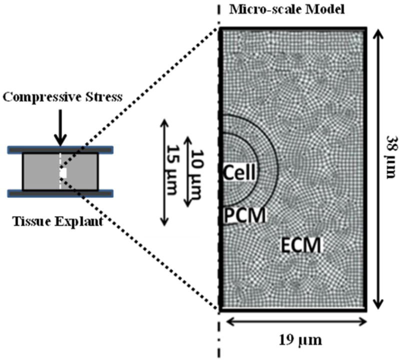
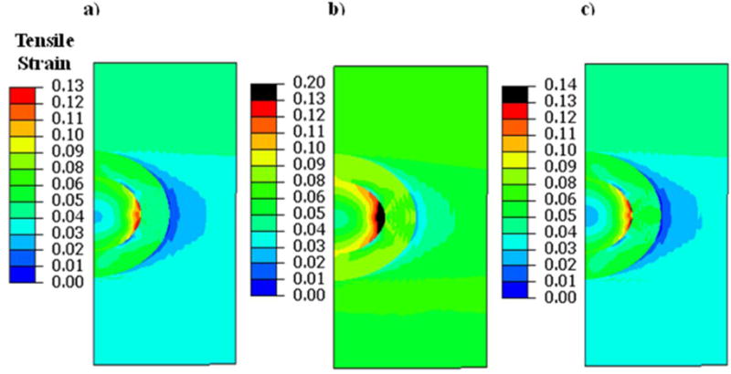
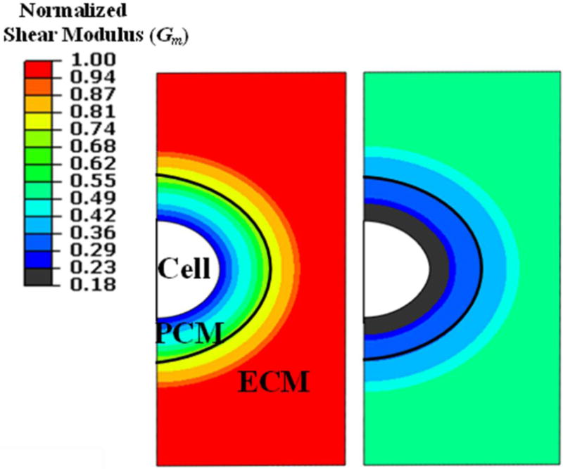
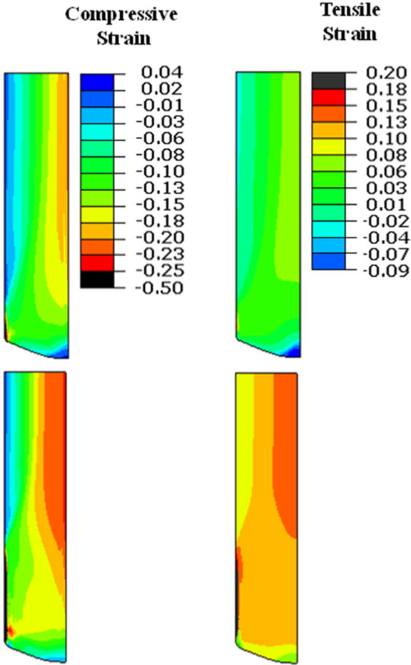
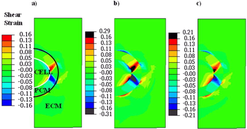
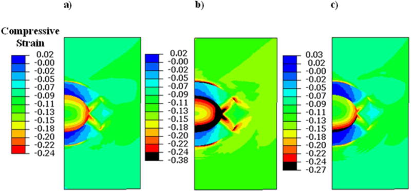
Understanding the mechanical factors that drive the biological responses of chondrocytes is central to our interpretation of the cascade of events that lead to osteoarthritic changes in articular cartilage. Chondrocyte mechanics is complicated by changes in tissue properties that can occur as osteoarthritis (OA) progresses and by the interaction between macro-scale, tissue level, properties, and micro-scale pericellular matrix (PCM) and local extracellular matrix (ECM) properties, both of which cannot be easily studied using in vitro systems. Our objective was to study the influence of macro- and micro-scale OA-associated structural changes on chondrocyte strains. We developed a multi-scale finite element model of articular cartilage subjected to unconfined loading, for the following three conditions: (i) normal articular cartilage, (ii) OA cartilage (where macro and micro-scale changes in collagen content, matrix modulus, and permeability were modeled), and (iii) early-stage OA cartilage (where only micro-scale changes in matrix modulus were modeled). In the macro-scale model, we found that a depth-dependent strain field was induced in both healthy and OA cartilage and that the middle and superficial zones of OA cartilage had increased tensile and compressive strains. At the micro-scale, chondrocyte shear strains were sensitive to PCM and local ECM properties. In the early-OA model, micro-scale spatial softening of PCM and ECM resulted in a substantial increase (30%) of chondrocyte shear strain, even with no structural changes in macro-scale tissue properties. Our study provides evidence that micromechanical changes at the cellular level may affect chondrocyte activities before macro-scale degradations at the tissue level become apparent. © 2017 Orthopaedic Research Society. Published by Wiley Periodicals, Inc. J Orthop Res 36:721-729, 2018.
了解驱动软骨细胞生物学反应的力学因素,对于我们解读导致关节软骨发生骨关节炎变化的一系列事件至关重要。随着骨关节炎(OA)的进展,组织特性会发生变化,同时宏观尺度的组织水平特性与微观尺度的细胞周围基质(PCM)和局部细胞外基质(ECM)特性之间存在相互作用,这使得软骨细胞力学变得复杂,而这两者都难以通过体外系统进行研究。我们的目标是研究宏观和微观尺度上与OA相关的结构变化对软骨细胞应变的影响。我们针对以下三种情况,开发了一个承受无侧限载荷的关节软骨多尺度有限元模型:(i)正常关节软骨,(ii)OA软骨(模拟了胶原含量、基质模量和渗透率在宏观和微观尺度上的变化),以及(iii)早期OA软骨(仅模拟了基质模量在微观尺度上的变化)。在宏观尺度模型中,我们发现健康软骨和OA软骨中均诱导出了深度依赖性应变场,且OA软骨的中层和表层区域的拉伸和压缩应变增加。在微观尺度上,软骨细胞剪应变对PCM和局部ECM特性敏感。在早期OA模型中,即使宏观尺度的组织特性没有结构变化,PCM和ECM在微观尺度上的空间软化也导致软骨细胞剪应变大幅增加(30%)。我们的研究提供了证据,表明在组织水平出现宏观降解之前,细胞水平的微机械变化可能会影响软骨细胞的活性。© 2017骨科研究协会。由威利期刊公司出版。《矫形外科学研究》36:721 - 729,2018年。