Kwong Tiffany C, Nouizi Farouk, Cho Jaedu, Lin Yuting, Sampathkumaran Uma, Gulsen Gultekin
Appl Opt. 2017 Oct 1;56(28):7886-7891. doi: 10.1364/AO.56.007886.
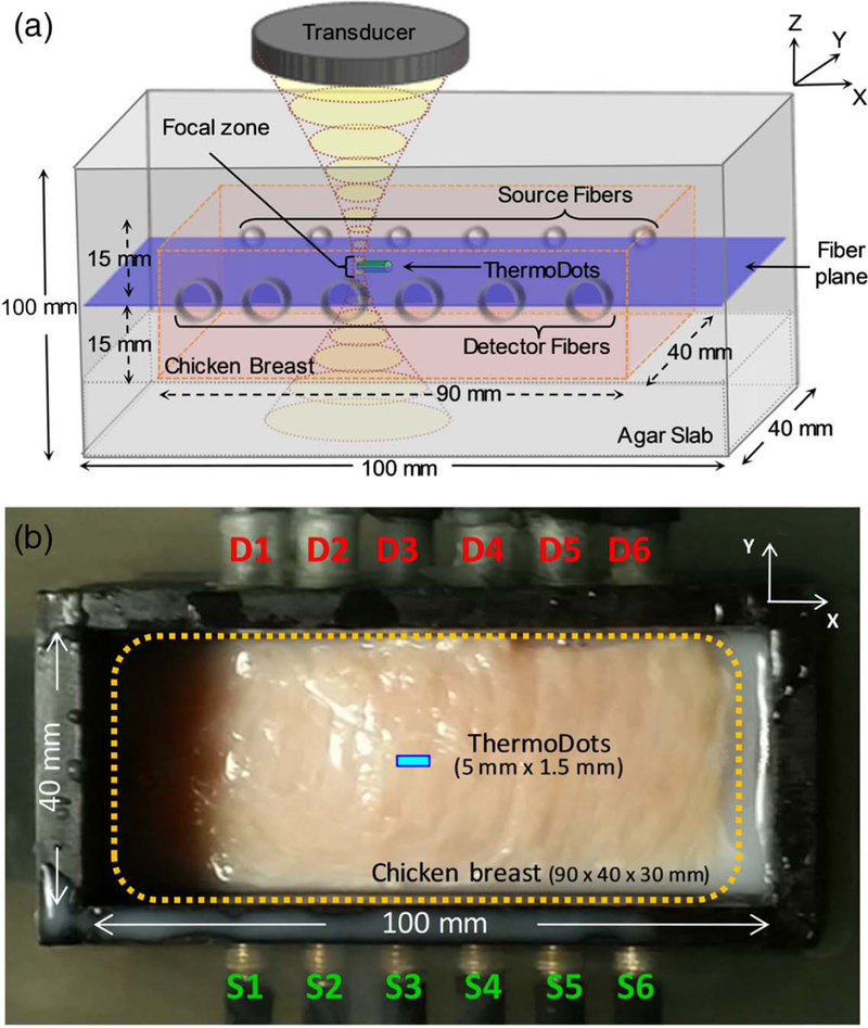
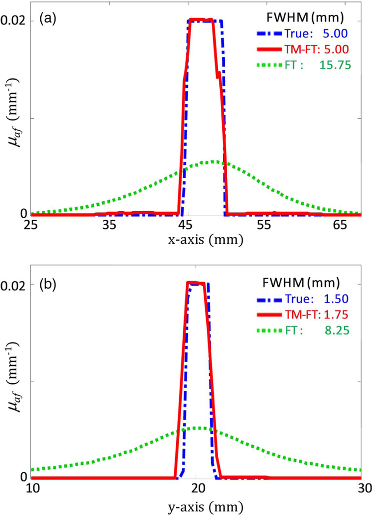
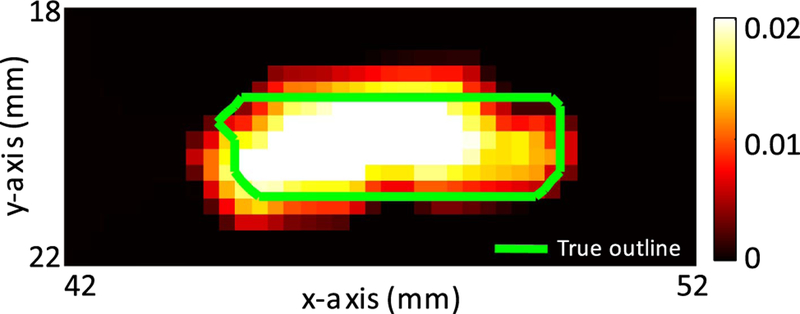
Previously, we demonstrated that temperature-modulated fluorescence tomography (TM-FT) could provide fluorescence images with high quantitative accuracy and the spatial resolution of focused ultrasound. TM-FT is based on scanning the focused ultrasound across the medium to activate temperature-reversible fluorescent nanoprobes (ThermoDots). This technique can resolve small fluorescent targets located several centimeters deep in turbid media with millimeter resolution. Our past studies with this multimodality technique used agar phantoms, which could not represent the true heterogeneous nature of the acoustic and optical properties of biological tissue. In this work, we report the results of the first TM-FT study performed on ex vivo chicken breast tissue. In order to improve the spatial resolution of this technique, diffuse optical tomography is also used to better estimate the optical property maps of the tissue, which is utilized as functional a priori for the TM-FT reconstruction algorithm. These ex vivo results show that TM-FT can accurately recover the concentration and position of a 1.5 mm×5 mm inclusion filled with ThermoDots. Since the inclusion is embedded 2 cm deep in the chicken breast sample, these results demonstrate the great potential of TM-FT for future in vivo small animal imaging.
此前,我们证明了温度调制荧光断层扫描(TM-FT)能够提供具有高定量准确性和聚焦超声空间分辨率的荧光图像。TM-FT基于在介质中扫描聚焦超声以激活温度可逆荧光纳米探针(热敏点)。该技术能够以毫米级分辨率分辨位于浑浊介质中几厘米深处的小荧光靶点。我们过去使用这种多模态技术进行的研究采用了琼脂体模,其无法体现生物组织声学和光学特性的真正异质性。在这项工作中,我们报告了首次在离体鸡胸组织上进行的TM-FT研究结果。为了提高该技术的空间分辨率,还使用了扩散光学断层扫描来更好地估计组织的光学特性图,并将其用作TM-FT重建算法的功能先验信息。这些离体结果表明,TM-FT能够准确恢复填充有热敏点的1.5毫米×5毫米内含物的浓度和位置。由于该内含物嵌入鸡胸样本2厘米深处,这些结果证明了TM-FT在未来小动物活体成像方面的巨大潜力。