Collaborative Mass Spectrometry Innovation Center, Skaggs School of Pharmacy and Pharmaceutical Sciences, University of California, San Diego, La Jolla, CA 92093, USA.
Department of Computer Science & Engineering, University of California, San Diego, La Jolla, CA 92093, USA.
Cell Host Microbe. 2017 Nov 8;22(5):705-716.e4. doi: 10.1016/j.chom.2017.10.001. Epub 2017 Oct 19.
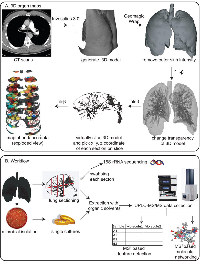
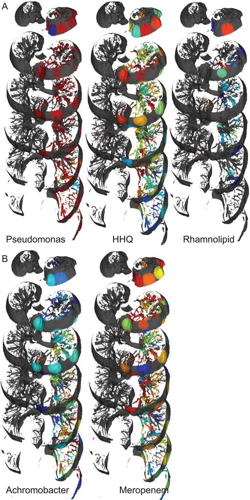
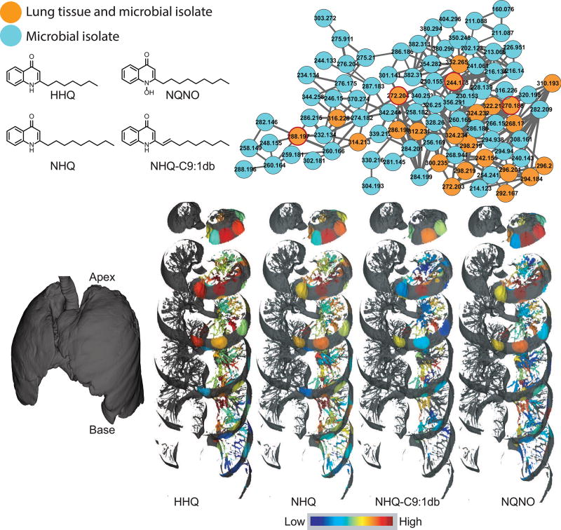
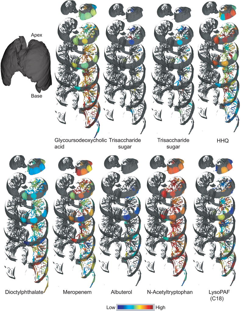
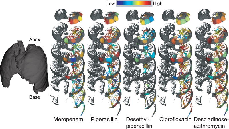
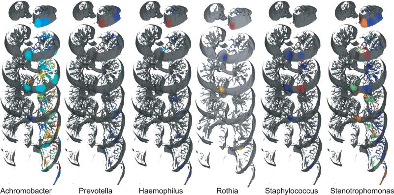
Our understanding of the spatial variation in the chemical and microbial makeup of an entire human organ remains limited, in part due to the size and heterogeneity of human organs and the complexity of the associated metabolome and microbiome. To address this challenge, we developed a workflow to enable the cartography of metabolomic and microbiome data onto a three-dimensional (3D) organ reconstruction built off radiological images. This enabled the direct visualization of the microbial and chemical makeup of a human lung from a cystic fibrosis patient. We detected host-derived molecules, microbial metabolites, medications, and region-specific metabolism of medications and placed it in the context of microbial distributions in the lung. Our tool further created browsable maps of a 3D microbiome/metabolome reconstruction map on a radiological image of a human lung and forms an interactive resource for the scientific community.
我们对整个人体器官的化学和微生物组成的空间变化的理解仍然有限,部分原因是人体器官的大小和异质性以及相关代谢组和微生物组的复杂性。为了应对这一挑战,我们开发了一种工作流程,能够将代谢组学和微生物组学数据映射到基于放射图像构建的三维(3D)器官重建上。这使得我们能够直接观察来自囊性纤维化患者的人类肺部的微生物和化学成分。我们检测到宿主来源的分子、微生物代谢物、药物以及药物在肺部的特定区域代谢,并将其置于肺部微生物分布的背景下。我们的工具进一步创建了人类肺部放射图像上的 3D 微生物组/代谢组重建图的可浏览地图,并为科学界形成了一个交互式资源。