Fox Grace E, Li Meng, Zhao Fang, Tsien Joe Z
Brain and Behavior Discovery Institute and Department of Neurology, Medical College of Georgia, Augusta University, Augusta, GA, United States of America.
The Brain Decoding Center, Banna Biomedical Research Institute, Xi-Shuang-Ban-Na Prefecture, Yunnan Province, China.
PLoS One. 2017 Oct 26;12(10):e0187198. doi: 10.1371/journal.pone.0187198. eCollection 2017.
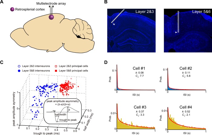
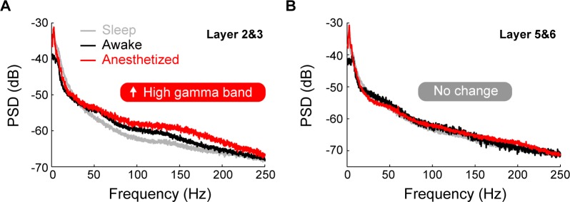
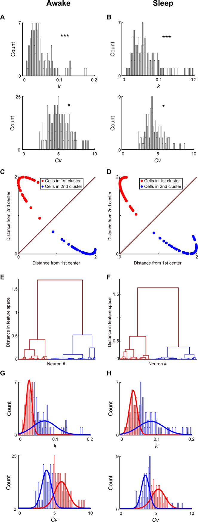
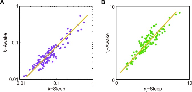
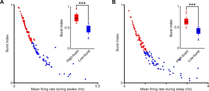
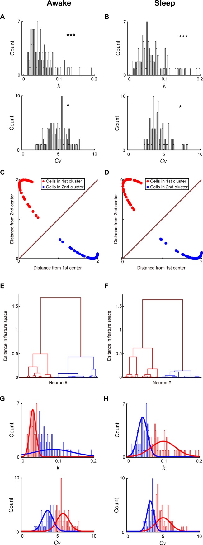
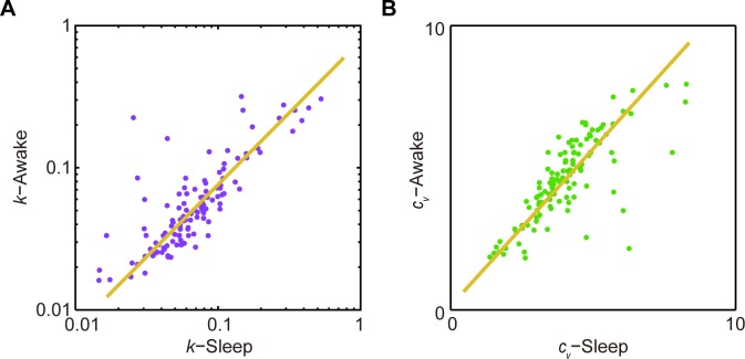
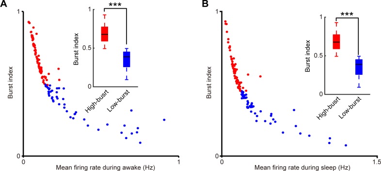
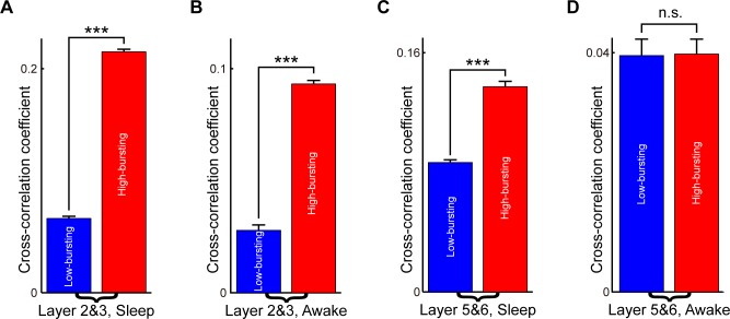
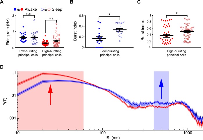
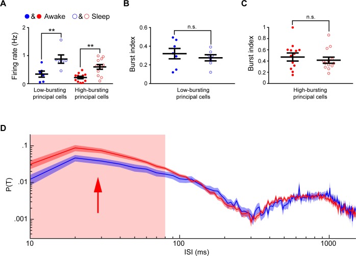
Ketamine is known to induce psychotic-like symptoms, including delirium and visual hallucinations. It also causes neuronal damage and cell death in the retrosplenial cortex (RSC), an area that is thought to be a part of high visual cortical pathways and at least partially responsible for ketamine's psychotomimetic activities. However, the basic physiological properties of RSC cells as well as their response to ketamine in vivo remained largely unexplored. Here, we combine a computational method, the Inter-Spike Interval Classification Analysis (ISICA), and in vivo recordings to uncover and profile excitatory cell subtypes within layers 2&3 and 5&6 of the RSC in mice within both conscious, sleep, and ketamine-induced unconscious states. We demonstrate two distinct excitatory principal cell sub-populations, namely, high-bursting excitatory principal cells and low-bursting excitatory principal cells, within layers 2&3, and show that this classification is robust over the conscious states, namely quiet awake, and natural unconscious sleep periods. Similarly, we provide evidence of high-bursting and low-bursting excitatory principal cell sub-populations within layers 5&6 that remained distinct during quiet awake and sleep states. We further examined how these subtypes are dynamically altered by ketamine. During ketamine-induced unconscious state, these distinct excitatory principal cell subtypes in both layer 2&3 and layer 5&6 exhibited distinct dynamics. We also uncovered different dynamics of local field potential under various brain states in layer 2&3 and layer 5&6. Interestingly, ketamine administration induced high gamma oscillations in layer 2&3 of the RSC, but not layer 5&6. Our results show that excitatory principal cells within RSC layers 2&3 and 5&6 contain multiple physiologically distinct sub-populations, and they are differentially affected by ketamine.
已知氯胺酮会诱发类似精神病的症状,包括谵妄和视幻觉。它还会导致脾后皮质(RSC)中的神经元损伤和细胞死亡,该区域被认为是高级视觉皮质通路的一部分,并且至少部分负责氯胺酮的拟精神病活性。然而,RSC细胞的基本生理特性及其在体内对氯胺酮的反应在很大程度上仍未得到探索。在这里,我们结合一种计算方法,即峰峰间隔分类分析(ISICA)和体内记录,来揭示和描绘小鼠RSC第2&3层和第5&6层内兴奋性细胞亚型在清醒、睡眠以及氯胺酮诱导的无意识状态下的情况。我们在第2&3层中证明了两个不同的兴奋性主细胞亚群,即高爆发性兴奋性主细胞和低爆发性兴奋性主细胞,并表明这种分类在清醒状态下,即安静清醒和自然无意识睡眠期是稳定的。同样,我们提供了证据表明在第5&6层中存在高爆发性和低爆发性兴奋性主细胞亚群,它们在安静清醒和睡眠状态下保持不同。我们进一步研究了这些亚型如何被氯胺酮动态改变。在氯胺酮诱导的无意识状态下,第2&3层和第5&6层中这些不同的兴奋性主细胞亚型表现出不同的动态变化。我们还发现了第2&3层和第5&6层在各种脑状态下局部场电位的不同动态变化。有趣 的是,给予氯胺酮会在RSC的第2&3层诱导高伽马振荡,但在第5&6层不会。我们的结果表明,RSC第2&3层和第5&6层内的兴奋性主细胞包含多个生理上不同的亚群,并且它们受到氯胺酮的影响不同。