Inserm, UMR 1069, Université François Rabelais Tours, Tours, France.
Network "Ion channels and cancer-Canceropole Grand Ouest, (IC-CGO), Grand Ouest, France.
Sci Rep. 2017 Oct 27;7(1):14199. doi: 10.1038/s41598-017-14230-1.
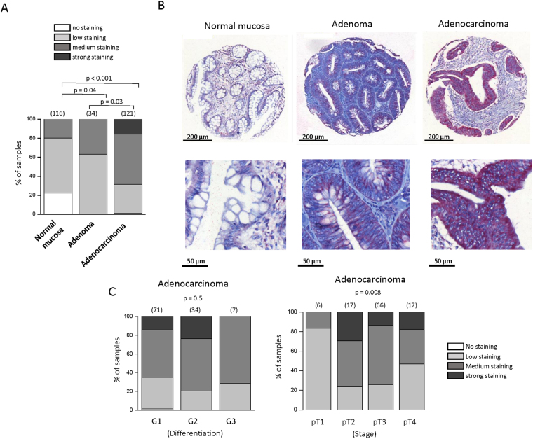
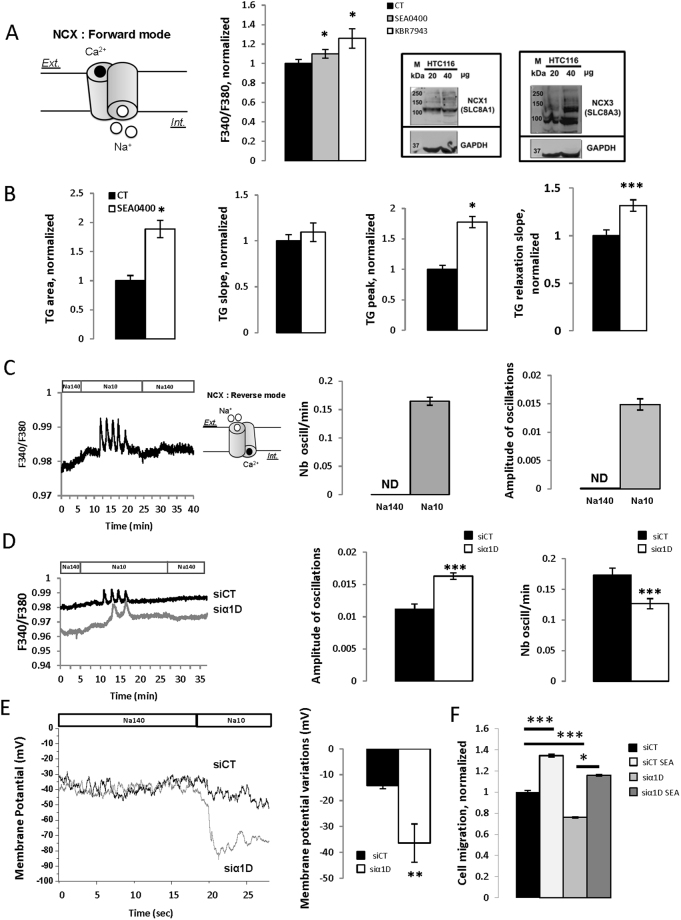
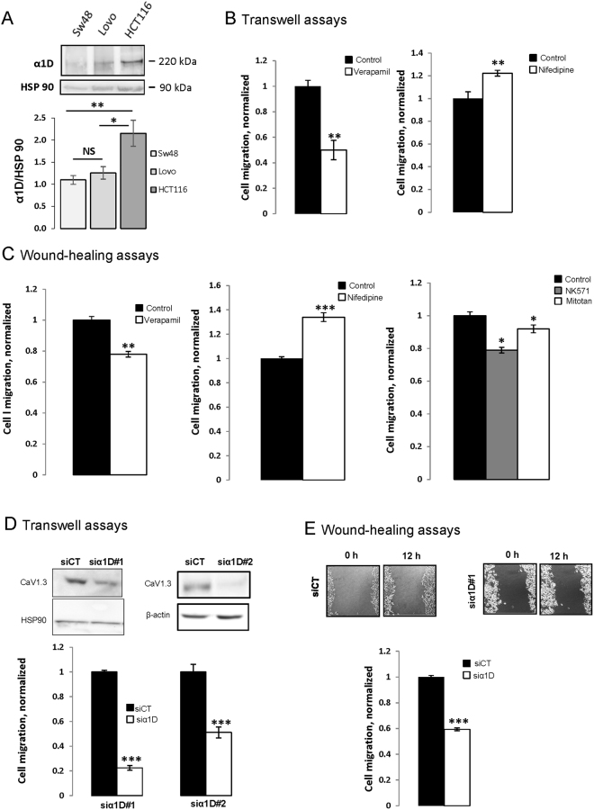
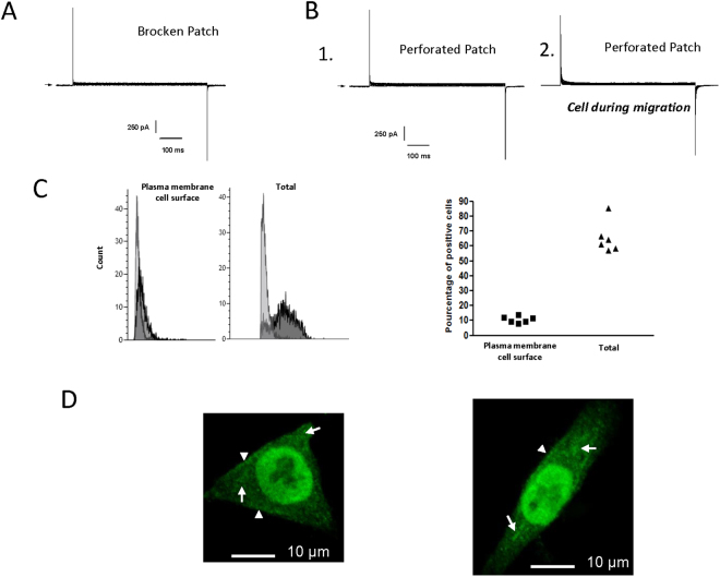
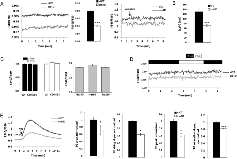
It is generally accepted that voltage-gated Ca channels, CaV, regulate Ca homeostasis in excitable cells following plasma membrane depolarization. Here, we show that the Ca protein α1D of CaV1.3 channel is overexpressed in colorectal cancer biopsies compared to normal tissues. Gene silencing experiments targeting α1D reduced the migration and the basal cytosolic Ca concentration of HCT116 colon cancer cell line and modified the cytosolic Ca oscillations induced by the sodium/calcium exchanger NCX1/3 working in its reverse mode. Interestingly, NCX1/3 regulated membrane potential of HCT116 cells only when α1D was silenced, and blocking NCX1/3 increased cytosolic Ca concentration and cell migration. However, membrane depolarization did not induce an increase in intracellular Ca. Patch-clamp experiments clearly showed that the inward Ca current was absent. Finally, flow cytometry and immunofluorescence studies showed that α1D protein was localized at the plasma membrane, in cytosol and cell nuclei. Altogether, we uncover a novel signaling pathway showing that α1D is involved in the regulation of Ca homeostasis and cell migration by a mechanism independent of its plasma membrane canonical function but that involved plasma membrane Na/Ca exchanger.
人们普遍认为,电压门控钙通道(CaV)在质膜去极化后调节兴奋细胞中的钙稳态。在这里,我们发现与正常组织相比,CaV1.3 通道的钙蛋白α1D在结直肠癌细胞活检中过度表达。针对α1D 的基因沉默实验降低了 HCT116 结肠癌细胞系的迁移和基础胞质 Ca 浓度,并改变了由钠/钙交换器 NCX1/3 以反向模式工作诱导的胞质 Ca 振荡。有趣的是,只有在沉默α1D 时,NCX1/3 才能调节 HCT116 细胞的膜电位,并且阻断 NCX1/3 会增加胞质 Ca 浓度和细胞迁移。然而,膜去极化不会引起细胞内 Ca 浓度增加。膜片钳实验清楚地表明不存在内向 Ca 电流。最后,流式细胞术和免疫荧光研究表明,α1D 蛋白定位于质膜、细胞质和细胞核。总之,我们揭示了一种新的信号通路,表明α1D 通过一种独立于其质膜经典功能但涉及质膜 Na/Ca 交换器的机制参与钙稳态和细胞迁移的调节。