Nasrin Sahela, Cader Fathima Aaysha, Haq M Maksumul, Karim Md Rezaul
Department of Cardiology, Ibrahim Cardiac Hospital & Research Institute (ICHRI), Dhaka, Bangladesh.
National Institute of Cardiovascular Diseases, Dhaka, Bangladesh.
BMC Res Notes. 2017 Oct 30;10(1):537. doi: 10.1186/s13104-017-2867-3.
Right coronary artery perforation extending to the sinus of Valsalva is a rare and potentially fatal complication of percutaneous coronary intervention. There are no definite guidelines on the management strategies for such complications. Treatment modality depends on the patient's haemodynamic stability and the extent of aortic involvement. Polytetrafluoroethylene-covered stents have emerged as a revolutionary strategy, enabling efficient endovascular repair of the entry port of such dissections, particularly the coronary ostia, and obviating the need for high-risk emergent surgical intervention.
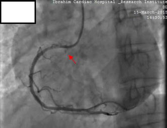
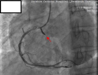
A 60 year old Bangladeshi gentleman underwent a coronary angiogram following a prior inferior ST elevation myocardial infarction (MI), 1 month previously. Coronary angiography done via right radial approach using 5 FR TIG catheter showed diffuse mid RCA disease with maximum 90% stenosis. Angioplasty of the RCA was planned. The RCA was cannulated with a 6-French JR 3.5 guiding catheter (USA). The lesion was crossed by a 0.014 inch guide wire and stented with a 2.75 × 38 mm novolimus-eluting DESyne stent, after predilatation. Immediately after stenting, a Type II perforation was observed in the ostial RCA, which progressed into the right coronary sinus of Valsalva. As the patient was haemodynamically stable with no ischaemia on ECG, we attempted to seal the ostial RCA with bare metal stents. Two successive bare metal stents failed to seal the aorto-coronary dissection. Ultimately, a 3.0 × 19 mm polytetrafluoroethylene-covered stent was deployed to seal the entry port in the ostial RCA, yielding a satisfactory angiographic result with only minimal contrast staining limited to the right sinus of Valsalva. The patient was closely monitored and discharged on dual antiplatelet therapy comprising of aspirin and prasugrel. He remained asymptomatic and with follow up echocardiograms showing no pericardial effusion nor extension of the dissection.
The polytetrafluoroethylene-covered stent provides a safe and effective means of sealing iatrogenic aorto-coronary dissections complicated by Ellis type II or II perforations, thus avoiding emergency surgery. However, as they are associated with increased incidence of stent thrombosis, an efficient and prolonged post-PCI antiplatelet regimen is recommended.
右冠状动脉穿孔延伸至主动脉窦是经皮冠状动脉介入治疗中一种罕见且可能致命的并发症。对于此类并发症的处理策略尚无明确的指南。治疗方式取决于患者的血流动力学稳定性以及主动脉受累的程度。聚四氟乙烯覆膜支架已成为一种革命性的策略,能够有效地对这类夹层的入口进行血管内修复,尤其是冠状动脉开口处,从而避免了高风险的急诊手术干预。
一名60岁的孟加拉国男性,在1个月前曾发生下壁ST段抬高型心肌梗死,随后接受了冠状动脉造影。通过右桡动脉途径使用5FR TIG导管进行的冠状动脉造影显示,右冠状动脉中段弥漫性病变,最大狭窄程度达90%。计划对右冠状动脉进行血管成形术。使用6F JR 3.5指引导管(美国)对右冠状动脉进行插管。病变部位通过一根0.014英寸的导丝穿过,并在预扩张后用一枚2.75×38mm的诺伐他汀洗脱DESyne支架进行支架植入。支架植入后立即观察到右冠状动脉开口处出现Ⅱ型穿孔,并进展至主动脉右窦。由于患者血流动力学稳定,心电图无缺血表现,我们尝试用裸金属支架封闭右冠状动脉开口。连续两枚裸金属支架未能封闭主动脉 - 冠状动脉夹层。最终,植入一枚3.0×19mm的聚四氟乙烯覆膜支架以封闭右冠状动脉开口处的入口,血管造影结果满意,仅在主动脉右窦有少量造影剂染色。对患者进行密切监测,并给予阿司匹林和普拉格雷组成的双联抗血小板治疗后出院。他一直无症状,随访超声心动图显示无心包积液,夹层也无扩展。
聚四氟乙烯覆膜支架为封闭并发EllisⅡ型或Ⅱ型穿孔的医源性主动脉 - 冠状动脉夹层提供了一种安全有效的方法,从而避免了急诊手术。然而,由于它们与支架血栓形成的发生率增加有关,建议采用高效且延长的PCI术后抗血小板治疗方案。