Lu Wu, Ke He, Qianshan Ding, Zhen Wang, Guoan Xiang, Honggang Yu
Department of Gastroenterology, Renmin Hospital of Wuhan University, Wuhan, China.
Hubei Key laboratory of Digestive System, Renmin Hospital of Wuhan University, Wuhan, China.
Iran J Basic Med Sci. 2017 Sep;20(9):990-995. doi: 10.22038/IJBMS.2017.9263.
Apatinib recently has been used to treat patients with gastric cancer, but the function of apatinib in colon cancer remains unclear. This study was conducted to investigate the impacts of apatinib on the biological function and its potential mechanism of colon cancer cells .
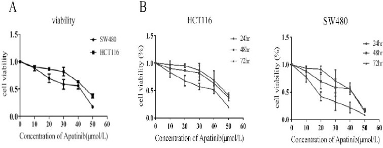
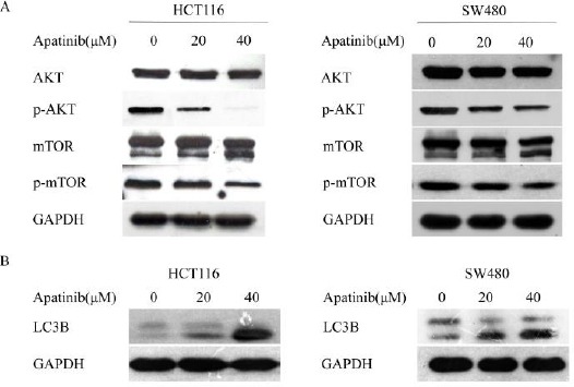
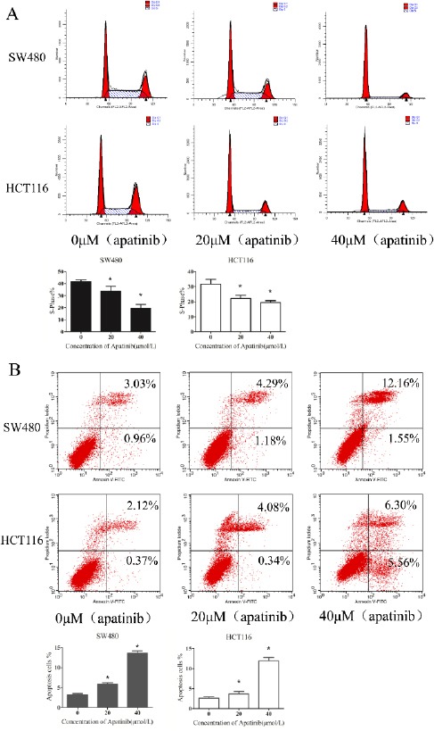
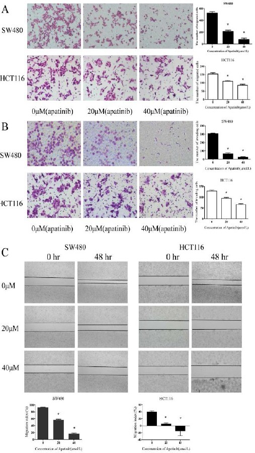
The effect of apatinib in colon cancer cells were detected by assessing cell viability, migration and invasion capabilities. Apoptosis cells and the cell cycle distribution of colon cancer cells were analyzed by flow cytometry. The potential mechanism was investigated via autophagy related proteins and pathways .
The proliferation, migration and invasion of colon cancer cells were inhibited when they were treated with different concentration of apatinib (20, 40 μM). When HCT116 and SW480 cells were treated with apatinib at the concentration of 20 μM, the apoptosis percentage were 3.7% and 5.8% respectively. As the drug concentration increased to 40μΜ, the the apoptosis percentage increased to 11.9% and 13.5%. Meanwhile, cell cycle was also altered. Furthermore, apatinib inhibited the expression of AKT-mTOR signaling pathway and increased the expression of LC3-II.
Apatinib can significantly inhibit the malignant phenotype of colon cancer cells, and it was involved in regulation of autophagy.
阿帕替尼最近已被用于治疗胃癌患者,但阿帕替尼在结肠癌中的作用仍不清楚。本研究旨在探讨阿帕替尼对结肠癌细胞生物学功能的影响及其潜在机制。
通过评估细胞活力、迁移和侵袭能力来检测阿帕替尼对结肠癌细胞的作用。采用流式细胞术分析结肠癌细胞的凋亡细胞和细胞周期分布。通过自噬相关蛋白和通路研究潜在机制。
用不同浓度(20、40 μM)的阿帕替尼处理结肠癌细胞时,其增殖、迁移和侵袭受到抑制。当用20 μM阿帕替尼处理HCT116和SW480细胞时,凋亡率分别为3.7%和5.8%。随着药物浓度增加到40 μM,凋亡率分别增加到11.9%和13.5%。同时,细胞周期也发生改变。此外,阿帕替尼抑制AKT-mTOR信号通路的表达并增加LC3-II的表达。
阿帕替尼可显著抑制结肠癌细胞的恶性表型,并参与自噬调节。