Ragusi M A A D, van der Meer R W, Joemai R M S, van Schaik J, van Rijswijk C S P
Department of Radiology, Leiden University Medical Center, Albinusdreef 2, P.O. Box 9600, 2300RC, Leiden, The Netherlands.
Department of Surgery, Leiden University Medical Center, Albinusdreef 2, P.O. Box 9600, 2300RC, Leiden, The Netherlands.
Cardiovasc Intervent Radiol. 2018 Feb;41(2):323-329. doi: 10.1007/s00270-017-1812-0. Epub 2017 Oct 30.
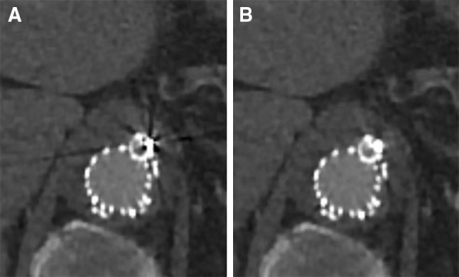
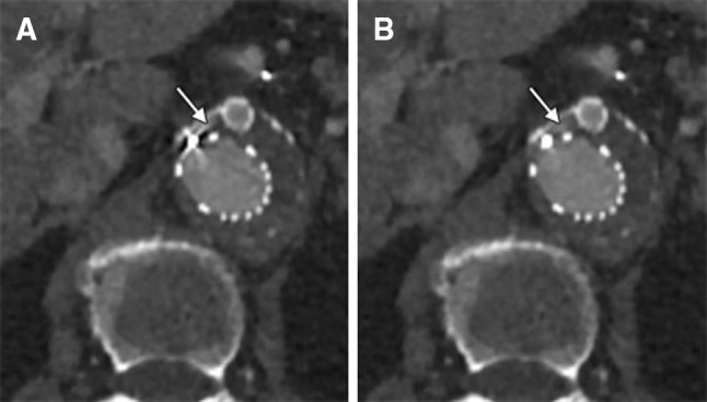
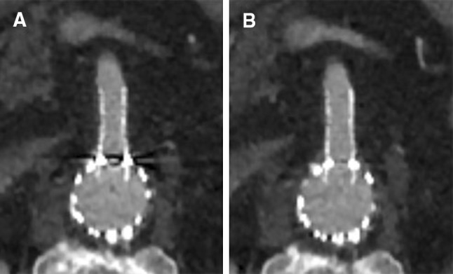
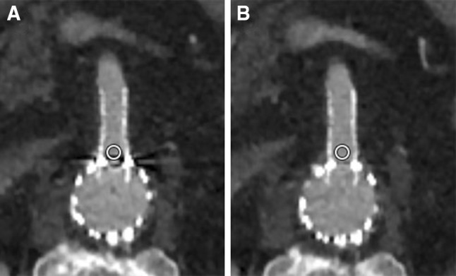
To evaluate the value of single-energy metal artifact reduction (SEMAR) algorithm on image quality in patients after complex endovascular aortic repair (EVAR) with fenestrated and branched devices.
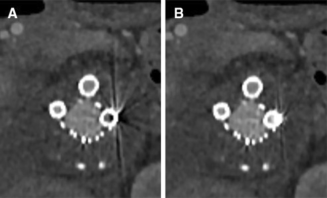
Routine follow-up computed tomography angiography (CTA) examinations were performed between February 2016 and May 2017 in 18 patients who underwent a complex EVAR procedure at our institution. Objective analysis was performed by measuring the standard deviation (SD) of attenuation (Hounsfield Units), and the contrast-to-noise ratio (CNR) in regions of interests in the stented visceral arteries. Subjective analysis of the degree of artifacts and stent visualization was performed independently by two interventional radiologists, blinded to the image reconstruction.
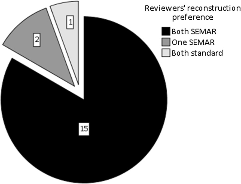
The SD of attenuation was significantly lower in all target visceral arteries (p < .001), the celiac artery (p = .002), the superior mesenteric artery (SMA; p = .043), and renal arteries (p < .001) in the CT images with SEMAR reconstruction. The CNR significantly increased in all SEMAR-reconstructed target visceral arteries (overall: p < .001, celiac artery: p = .009; SMA: p = .003; renal arteries: p < .001). The reviewers rated a significantly lower artifact degree in all target vessels (overall: p < .001, celiac artery: p = .001; SMA: p = .008; renal arteries: p < .001) and a significantly improved visualization of the stent patency in all target vessels (overall: p < .001, celiac artery: p = .031; SMA: p = .047; renal arteries: p < .001) in the SEMAR images. Overall preference of both reviewers was in favor of the SEMAR reconstruction in 15/18 cases (83%).
Reconstruction with SEMAR algorithm significantly improves CTA image quality in patients after complex EVAR.
Level 4, Case series.
评估单能量金属伪影减少(SEMAR)算法对接受带开窗和分支装置的复杂血管腔内主动脉修复术(EVAR)患者图像质量的影响。
2016年2月至2017年5月期间,对在我院接受复杂EVAR手术的18例患者进行常规随访计算机断层扫描血管造影(CTA)检查。通过测量支架置入的内脏动脉感兴趣区域的衰减标准差(SD,亨氏单位)和对比噪声比(CNR)进行客观分析。由两名介入放射科医生独立对伪影程度和支架可视化程度进行主观分析,他们对图像重建情况不知情。
在采用SEMAR重建的CT图像中,所有目标内脏动脉(p <.001)、腹腔动脉(p =.002)、肠系膜上动脉(SMA;p =.043)和肾动脉(p <.001)的衰减SD均显著降低。在所有采用SEMAR重建的目标内脏动脉中,CNR显著升高(总体:p <.001,腹腔动脉:p =.009;SMA:p =.003;肾动脉:p <.001)。 reviewers对所有目标血管的伪影程度评分显著降低(总体:p <.001,腹腔动脉:p =.001;SMA:p =.008;肾动脉:p <.001),且对所有目标血管中支架通畅情况的可视化程度显著改善(总体:p <.001,腹腔动脉:p =.031;SMA:p =.047;肾动脉:p <.001)。两位reviewers的总体偏好是在15/18例(83%)病例中支持SEMAR重建。
采用SEMAR算法重建可显著提高复杂EVAR术后患者的CTA图像质量。
4级,病例系列。