Klejewski Andrzej, Sterzyńska Karolina, Wojtowicz Karolina, Świerczewska Monika, Partyka Małgorzata, Brązert Maciej, Nowicki Michał, Zabel Maciej, Januchowski Radosław
Department of Nursing, Poznań University of Medical Sciences, Poznań, Poland.
Department of Obstetrics and Womens Diseases, Poznań University of Medical Sciences, Poznań, Poland.
Oncotarget. 2017 Aug 10;8(43):74466-74478. doi: 10.18632/oncotarget.20169. eCollection 2017 Sep 26.
The aim of the present study is to determine the expression of LUM in drug-resistant ovarian cancer cell lines.
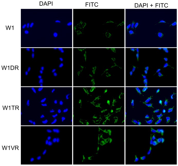
Doxorubicin- (DOX), topotecan- (TOP) and vincristine- (VIN) resistant variants of the W1 ovarian cancer cell line were used in this study. We used quantitative real-time polymerase chain reactions to determine LUM mRNA levels. Protein expression was detected using Western blot and immunocytochemistry assays. Protein glycosylation was investigated using PGNase F digestion. Immunohistochemistry assays were used to determine protein expression in ovarian cancer patients.
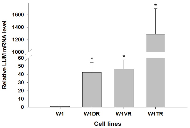
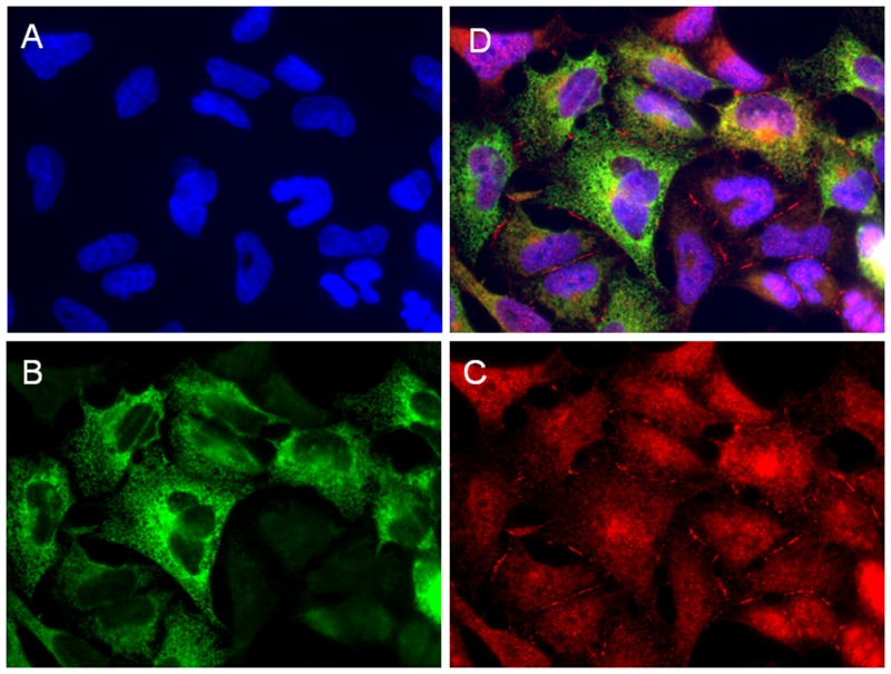
We observed increased expression of LUM in drug-resistant cell lines at both the mRNA and the protein level. The most abundant LUM expression was observed in TOP-resistant cell line. We observed LUM bands that corresponded to different molecular masses, and the most abundant LUM form was the secreted form, which had a mass of 50 kDa. Double immunofluorescence analysis showed co-expression of LUM and COL3A1 as well as the presence of extracellular COL3A1 in the TOP-resistant cell line. Finally, we detected the LUM protein in ovarian cancer tissue.
The expression of LUM in cytostatic-resistant cell lines suggests its role in drug resistance. The co-expression of LUM and COL3A1 indicates the significance of LUM in collagen fibre assembly. Expression in ovarian cancer tissue suggests that LUM can play a role in ovarian cancer pathogenesis in ways similar to other cancers.
本研究旨在确定LUM在耐药性卵巢癌细胞系中的表达情况。
本研究使用了W1卵巢癌细胞系的阿霉素(DOX)、拓扑替康(TOP)和长春新碱(VIN)耐药变体。我们采用定量实时聚合酶链反应来确定LUM mRNA水平。使用蛋白质印迹法和免疫细胞化学分析法检测蛋白质表达。使用PNG酶F消化法研究蛋白质糖基化。采用免疫组织化学分析法确定卵巢癌患者中的蛋白质表达。
我们观察到耐药细胞系中LUM在mRNA和蛋白质水平上的表达均增加。在TOP耐药细胞系中观察到最丰富的LUM表达。我们观察到与不同分子量相对应的LUM条带,最丰富的LUM形式是分泌形式,其分子量为50 kDa。双重免疫荧光分析显示在TOP耐药细胞系中LUM和COL3A1共表达以及细胞外COL3A1的存在。最后,我们在卵巢癌组织中检测到了LUM蛋白。
LUM在细胞生长抑制剂耐药细胞系中的表达表明其在耐药性中发挥作用。LUM和COL3A1的共表达表明LUM在胶原纤维组装中的重要性。在卵巢癌组织中的表达表明LUM可能以与其他癌症相似的方式在卵巢癌发病机制中发挥作用。