• 文献检索
  • 文档翻译
  • 深度研究
  • 学术资讯
  • Suppr Zotero 插件Zotero 插件
  • 邀请有礼
  • 套餐&价格
  • 历史记录
应用&插件
Suppr Zotero 插件Zotero 插件浏览器插件Mac 客户端Windows 客户端微信小程序
定价
高级版会员购买积分包购买API积分包
服务
文献检索文档翻译深度研究API 文档MCP 服务
关于我们
关于 Suppr公司介绍联系我们用户协议隐私条款
关注我们

Suppr 超能文献

核心技术专利:CN118964589B侵权必究
粤ICP备2023148730 号-1Suppr @ 2026

文献检索

告别复杂PubMed语法,用中文像聊天一样搜索,搜遍4000万医学文献。AI智能推荐,让科研检索更轻松。

立即免费搜索

文件翻译

保留排版,准确专业,支持PDF/Word/PPT等文件格式,支持 12+语言互译。

免费翻译文档

深度研究

AI帮你快速写综述,25分钟生成高质量综述,智能提取关键信息,辅助科研写作。

立即免费体验

李斯特结节的解剖变异:基于磁共振成像的一种新的形态学分类

Anatomical Variants of Lister's Tubercle: A New Morphological Classification Based on Magnetic Resonance Imaging.

作者信息

Chan Wan Ying, Chong Le Roy

机构信息

Department of Radiology, Changi General Hospital, Singapore 529889, Singapore.

出版信息

Korean J Radiol. 2017 Nov-Dec;18(6):957-963. doi: 10.3348/kjr.2017.18.6.957. Epub 2017 Sep 21.

DOI:10.3348/kjr.2017.18.6.957
PMID:29089828
原文链接:https://pmc.ncbi.nlm.nih.gov/articles/PMC5639161/
Abstract

OBJECTIVE

Lister's tubercle is used as a standard anatomical landmark in hand surgery and arthroscopy procedures. In this study, we aimed to evaluate and propose a classification for anatomical variants of Lister's tubercle.

MATERIALS AND METHODS

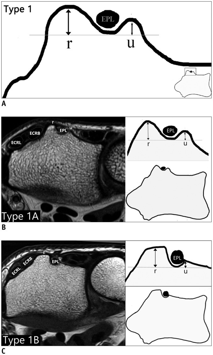
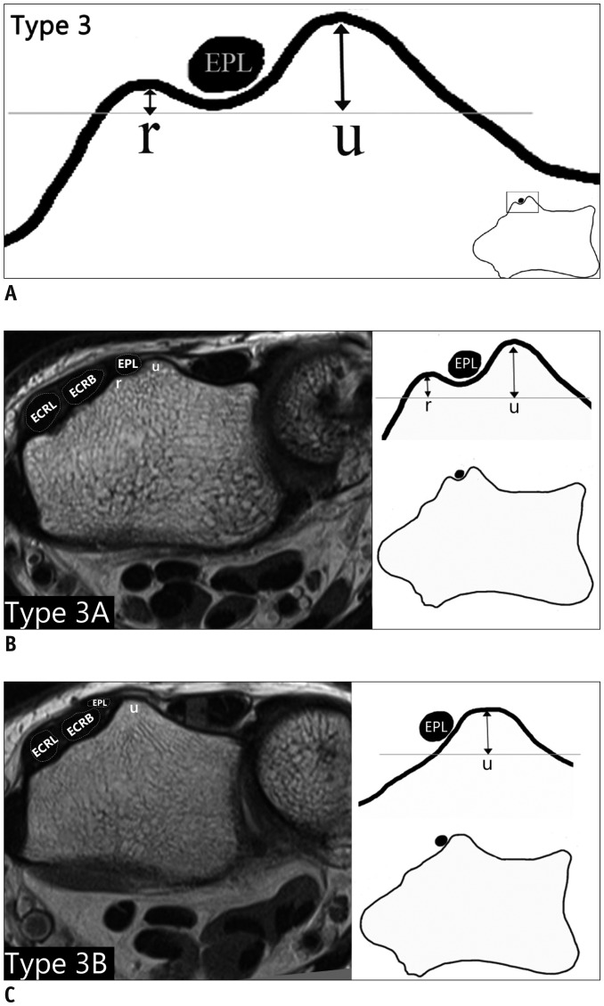
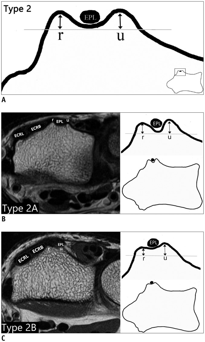
Between September 2011 and July 2014, 360 MRI examinations for wrists performed using 1.5T scanners in a single institution were retrospectively evaluated. The prevalence of anatomical variants of Lister's tubercle based on the heights and morphology of its radial and ulnar peaks was assessed. These were classified into three distinct types: radial peak larger than ulnar peak (Type 1), similar radial and ulnar peaks (Type 2) and ulnar peak larger than radial peak (Type 3). Each type was further divided into 2 subtypes (A and B) based on the morphology of the peaks.

RESULTS

The proportions of Type 1, Type 2, and Type 3 variants in the study population were 69.2, 21.4, and 9.5%, respectively. For the subtypes, the Type 1A variant was the most common (41.4%) and conformed to the classical appearance of Lister's tubercle; whereas, Type 3A and 3B variants were rare configurations (6.4% and 3.1%, respectively) wherein the extensor pollicis longus tendon coursed along the radial aspect of Lister's tubercle.

CONCLUSION

Anatomical variations of Lister's tubercle have potential clinical implications for certain pathological conditions and pre-procedural planning. The proposed classification system facilitates a better understanding of these anatomical variations and easier identification of at-risk and rare variants.

摘要

目的

在手部手术和关节镜手术中,李斯特结节用作标准解剖标志。在本研究中,我们旨在评估并提出李斯特结节解剖变异的分类。

材料与方法

回顾性评估2011年9月至2014年7月间在单一机构使用1.5T扫描仪对腕部进行的360例MRI检查。根据其桡侧和尺侧峰的高度和形态评估李斯特结节解剖变异的发生率。这些变异分为三种不同类型:桡侧峰大于尺侧峰(1型)、桡侧和尺侧峰相似(2型)以及尺侧峰大于桡侧峰(3型)。根据峰的形态,每种类型进一步分为2个亚型(A和B)。

结果

研究人群中1型、2型和3型变异的比例分别为69.2%、21.4%和9.5%。对于各亚型,1A型变异最为常见(41.4%),符合李斯特结节的经典外观;而3A型和3B型变异是罕见的形态(分别为6.4%和3.1%),其中拇长伸肌腱沿李斯特结节的桡侧走行。

结论

李斯特结节的解剖变异对某些病理状况和术前规划具有潜在临床意义。所提出的分类系统有助于更好地理解这些解剖变异,并更容易识别有风险和罕见的变异。

https://cdn.ncbi.nlm.nih.gov/pmc/blobs/54cc/5639161/f811f2a8ded2/kjr-18-957-g003.jpg
https://cdn.ncbi.nlm.nih.gov/pmc/blobs/54cc/5639161/cacdf31aa30c/kjr-18-957-g001.jpg
https://cdn.ncbi.nlm.nih.gov/pmc/blobs/54cc/5639161/a02f4645d833/kjr-18-957-g002.jpg
https://cdn.ncbi.nlm.nih.gov/pmc/blobs/54cc/5639161/f811f2a8ded2/kjr-18-957-g003.jpg
https://cdn.ncbi.nlm.nih.gov/pmc/blobs/54cc/5639161/cacdf31aa30c/kjr-18-957-g001.jpg
https://cdn.ncbi.nlm.nih.gov/pmc/blobs/54cc/5639161/a02f4645d833/kjr-18-957-g002.jpg
https://cdn.ncbi.nlm.nih.gov/pmc/blobs/54cc/5639161/f811f2a8ded2/kjr-18-957-g003.jpg

相似文献

1
Anatomical Variants of Lister's Tubercle: A New Morphological Classification Based on Magnetic Resonance Imaging.李斯特结节的解剖变异:基于磁共振成像的一种新的形态学分类
Korean J Radiol. 2017 Nov-Dec;18(6):957-963. doi: 10.3348/kjr.2017.18.6.957. Epub 2017 Sep 21.
2
Anatomical Localization of Lister's Tubercle and its Clinical and Surgical Importance.李斯特结节的解剖定位及其临床和外科意义。
Open Orthop J. 2014 Apr 4;8:74-7. doi: 10.2174/1874325001408010074. eCollection 2014.
3
Ultrasound assessment of extensor pollicis longus tendon rupture following distal radius fracture: a sonographic and surgical correlation.桡骨远端骨折后拇长伸肌腱断裂的超声评估:超声与手术的相关性
J Ultrason. 2020;20(80):e1-e5. doi: 10.15557/JoU.2020.0001. Epub 2020 Mar 31.
4
Anatomic Landmarks of the Distal Radioulnar Joint.桡尺远侧关节的解剖标志
Eplasty. 2015 Aug 14;15:e36. eCollection 2015.
5
Distal radial and ulnar landmarks used in percutaneous pin fixation: anatomical relationship to the superficial radial and ulnar nerves.经皮穿针固定术中使用的桡骨远端和尺骨远端标志:与桡神经浅支和尺神经浅支的解剖关系。
Hand Surg. 2010;15(3):161-4. doi: 10.1142/S0218810410004783.
6
Anatomic location of the first dorsal extensor compartment for surgical De-Quervain's tenosynovitis release: A cadaveric study.手术治疗桡骨茎突狭窄性腱鞘炎时第一背侧伸肌间隔的解剖位置:一项尸体研究
World J Orthop. 2024 Apr 18;15(4):379-385. doi: 10.5312/wjo.v15.i4.379.
7
Morphometric analysis of lister's tubercle and its consequences on volar plate fixation of distal radius fractures.李斯特结节的形态学分析及其对桡骨远端骨折掌侧板固定的影响。
J Hand Surg Am. 2008 Dec;33(10):1716-9. doi: 10.1016/j.jhsa.2008.08.012.
8
Anatomic surface landmarks to guide injection for posterior interosseous nerve block.指导骨间后神经阻滞注射的解剖表面标志。
J Plast Surg Hand Surg. 2021 Feb;55(1):17-20. doi: 10.1080/2000656X.2020.1828895. Epub 2020 Oct 10.
9
The anatomy of the posterior interosseous nerve as a graft.作为移植物的骨间后神经的解剖结构。
J Hand Surg Am. 2000 Sep;25(5):930-5. doi: 10.1053/jhsu.2000.16359.
10
A possible mechanism of direct injury to the EPL tendon at Lister's tubercle during falls with the wrist fully extended.在手腕完全伸展的跌倒过程中,EPL肌腱在Lister结节处受到直接损伤的一种可能机制。
J Hand Surg Eur Vol. 2008 Apr;33(2):149-51. doi: 10.1177/1753193407087575.

引用本文的文献

1
The Intraoperative Dorsal Tangential and the Radial Groove View of the Distal Radius to Detect Dorsal Screw Protrusion in Volar Plating: Teaching Effect and Value in Clinical Practice.术中桡骨远端背侧切线位及桡侧沟位用于检测掌侧钢板固定时背侧螺钉突出:教学效果及临床应用价值
J Hand Surg Glob Online. 2025 May 21;7(4):100734. doi: 10.1016/j.jhsg.2025.100734. eCollection 2025 Jul.
2
Osteometric Study of the Dorsal (Lister's) Tubercle of the Radius in Relation to the Neighboring Anatomical Elements: Suprastyloid, Accessory, and Oblique Crests.关于桡骨背侧(李斯特氏)结节与相邻解剖结构(上髁、副嵴和斜嵴)关系的骨测量学研究。
Life (Basel). 2025 Feb 11;15(2):273. doi: 10.3390/life15020273.
3

本文引用的文献

1
The Effectiveness of Corticosteroid Injection for De Quervain's Stenosing Tenosynovitis (DQST): A Systematic Review and Meta-Analysis.皮质类固醇注射治疗桡骨茎突狭窄性腱鞘炎(DQST)的有效性:一项系统评价和荟萃分析。
Open Orthop J. 2015 Sep 30;9:437-44. doi: 10.2174/1874325001509010437. eCollection 2015.
2
Anatomical Localization of Lister's Tubercle and its Clinical and Surgical Importance.李斯特结节的解剖定位及其临床和外科意义。
Open Orthop J. 2014 Apr 4;8:74-7. doi: 10.2174/1874325001408010074. eCollection 2014.
3
Radioscapholunate arthrodesis using low-profile dorsal pi plate.
Anatomy of Lister's Tubercle: Implications for Volar Locked Plating of the Distal Radius.
李斯特结节的解剖结构:对桡骨远端掌侧锁定钢板固定的意义
Hand (N Y). 2024 Oct 29:15589447241284783. doi: 10.1177/15589447241284783.
4
Spontaneous Rupture of the Extensor Pollicis Longus in a Semi-professional Bodybuilder.一名半职业健美运动员的拇长伸肌自发性断裂
Cureus. 2023 Feb 16;15(2):e35062. doi: 10.7759/cureus.35062. eCollection 2023 Feb.
5
Ultrasound assessment of extensor pollicis longus tendon rupture following distal radius fracture: a sonographic and surgical correlation.桡骨远端骨折后拇长伸肌腱断裂的超声评估:超声与手术的相关性
J Ultrason. 2020;20(80):e1-e5. doi: 10.15557/JoU.2020.0001. Epub 2020 Mar 31.
6
Age of Data in Contemporary Research Articles Published in Representative General Radiology Journals.当代代表性综合放射学期刊研究文章中的数据时代。
Korean J Radiol. 2018 Nov-Dec;19(6):1172-1178. doi: 10.3348/kjr.2018.19.6.1172. Epub 2018 Oct 18.
使用低轮廓背侧π形钢板的桡舟月关节融合术。
Tech Hand Up Extrem Surg. 2013 Jun;17(2):80-3. doi: 10.1097/BTH.0b013e318280e9cc.
4
Dorsal tangential view of the wrist to detect screw penetration to the dorsal cortex of the distal radius after volar fixed-angle plating.腕关节背侧切线位视图,用于检测掌侧锁定钢板固定后螺钉穿入桡骨远端背侧皮质的情况。
Hand (N Y). 2011 Jun;6(2):190-3. doi: 10.1007/s11552-010-9316-2. Epub 2011 Jan 7.
5
Anatomical relations of the superficial sensory branches of the radial nerve: a cadaveric study with clinical implications.桡神经浅感觉支的解剖关系:一项具有临床意义的尸体研究
Patient Saf Surg. 2011 Nov 4;5(1):28. doi: 10.1186/1754-9493-5-28.
6
Sonographic appearance of the posterior interosseous nerve at the wrist.腕关节骨间后神经的超声表现。
J Ultrasound Med. 2011 Sep;30(9):1233-9. doi: 10.7863/jum.2011.30.9.1233.
7
[Fixation of fractures of the distal radius using the "nail-plate"].使用“钉板”固定桡骨远端骨折
Oper Orthop Traumatol. 2009 Nov;21(4-5):459-71. doi: 10.1007/s00064-009-1912-3.
8
Computer tomography aided 3D analysis of the distal dorsal radius surface and the effects on volar plate osteosynthesis.计算机断层扫描辅助对桡骨远端背侧表面进行三维分析及其对掌侧板接骨术的影响
J Hand Surg Eur Vol. 2009 Oct;34(5):598-602. doi: 10.1177/1753193409101471.
9
Intraosseous vascularity of the distal radius: anatomy and clinical implications in distal radius fractures.桡骨远端的骨内血管:桡骨远端骨折的解剖学及临床意义
Hand (N Y). 2009 Dec;4(4):418-23. doi: 10.1007/s11552-009-9204-9. Epub 2009 May 28.
10
Anatomic landmarks for the first dorsal compartment.第一背侧间隔的解剖标志。
Eplasty. 2008;8:e53. Epub 2008 Nov 18.