Togo Tatsuru
Department of Anatomy, St. Marianna University School of Medicine, 2-16-1 Sugao, Miyamae, Kawasaki, Kanagawa 216-8511, Japan
Biol Open. 2017 Dec 15;6(12):1814-1819. doi: 10.1242/bio.028977.
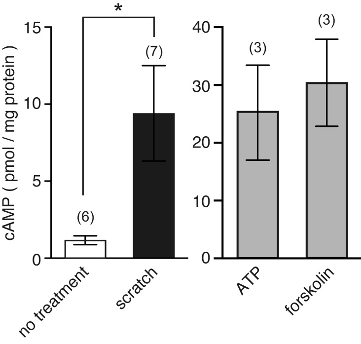
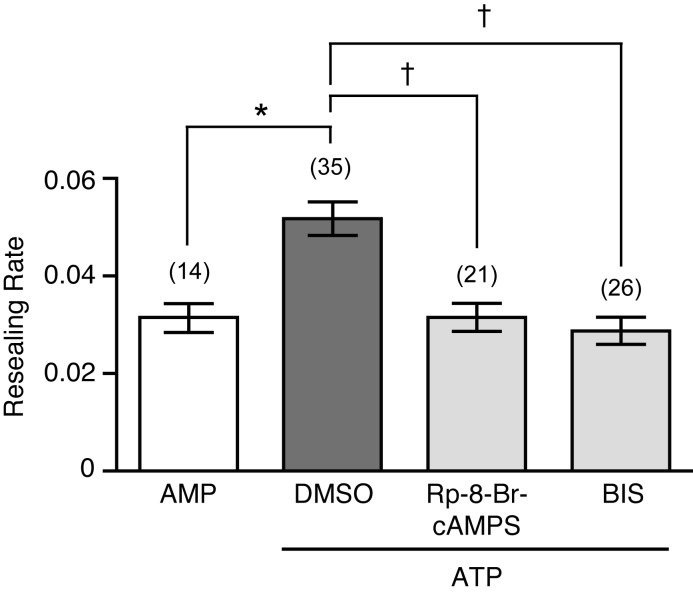
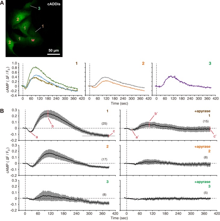
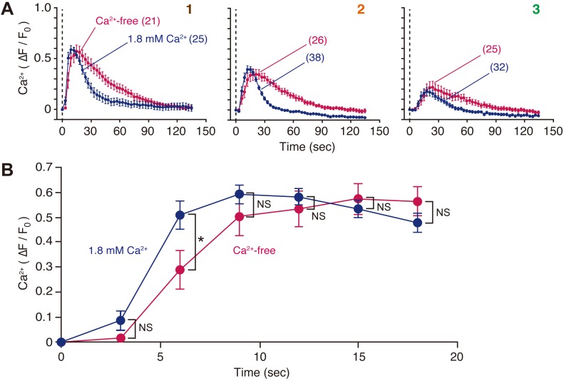
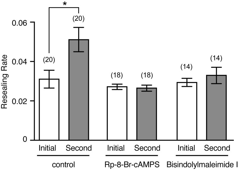
Disruption of cellular plasma membranes is a common event in many animal tissues, and the membranes are usually rapidly resealed. Moreover, repeated membrane disruptions within a single cell reseal faster than the initial wound in a protein kinase A (PKA)- and protein kinase C (PKC)-dependent manner. In addition to wounded cells, recent studies have demonstrated that wounding of Madin-Darby canine kidney (MDCK) cells potentiates membrane resealing in neighboring cells in the short-term by purinergic signaling, and in the long-term by nitric oxide/protein kinase G signaling. In the present study, real-time imaging showed that cell membrane disruption stimulated cAMP synthesis and Ca mobilization from intracellular stores by purinergic signaling in neighboring MDCK cells. Furthermore, inhibition of PKA and PKC suppressed the ATP-mediated short-term potentiation of membrane resealing in neighboring cells. These results suggest that cell membrane disruption stimulates PKA and PKC via purinergic signaling to potentiate cell membrane resealing in neighboring MDCK cells.
细胞膜破裂在许多动物组织中是常见现象,并且细胞膜通常会迅速重新封闭。此外,单个细胞内的反复膜破裂比初始伤口重新封闭得更快,这一过程依赖蛋白激酶A(PKA)和蛋白激酶C(PKC)。除了受伤细胞外,最近的研究表明,Madin-Darby犬肾(MDCK)细胞受伤后,在短期内通过嘌呤能信号传导增强相邻细胞的膜重新封闭,在长期内通过一氧化氮/蛋白激酶G信号传导增强相邻细胞的膜重新封闭。在本研究中,实时成像显示,细胞膜破裂通过嘌呤能信号传导刺激相邻MDCK细胞中的cAMP合成和从细胞内储存库释放钙。此外,抑制PKA和PKC可抑制ATP介导的相邻细胞中膜重新封闭的短期增强。这些结果表明,细胞膜破裂通过嘌呤能信号传导刺激PKA和PKC,以增强相邻MDCK细胞中的细胞膜重新封闭。