Sugiyama Atsuhiko, Sone Daichi, Sato Noriko, Kimura Yukio, Ota Miho, Maikusa Norihide, Maekawa Tomoko, Enokizono Mikako, Mori-Yoshimura Madoka, Ohya Yasushi, Kuwabara Satoshi, Matsuda Hiroshi
Department of Radiology, National Center of Neurology and Psychiatry, Tokyo, Japan.
Department of Neurology, Graduate School of Medicine, Chiba University, Chiba, Japan.
PLoS One. 2017 Nov 2;12(11):e0187343. doi: 10.1371/journal.pone.0187343. eCollection 2017.
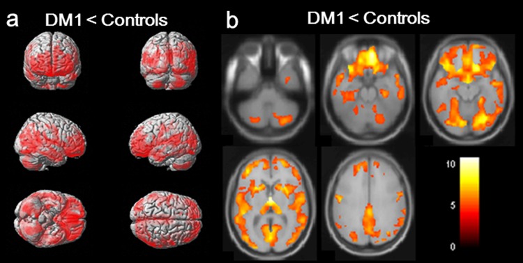
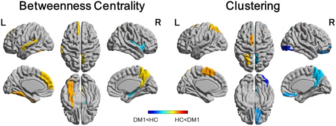
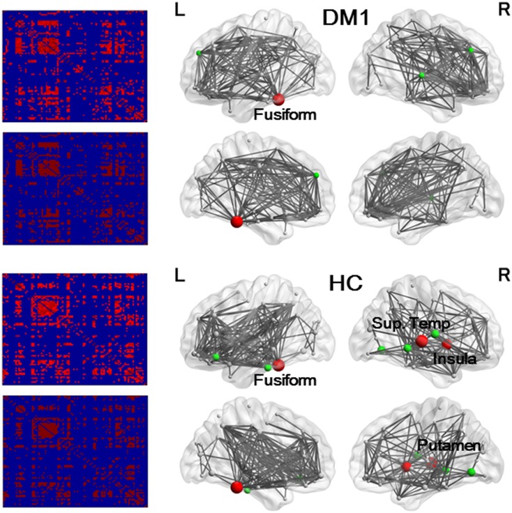
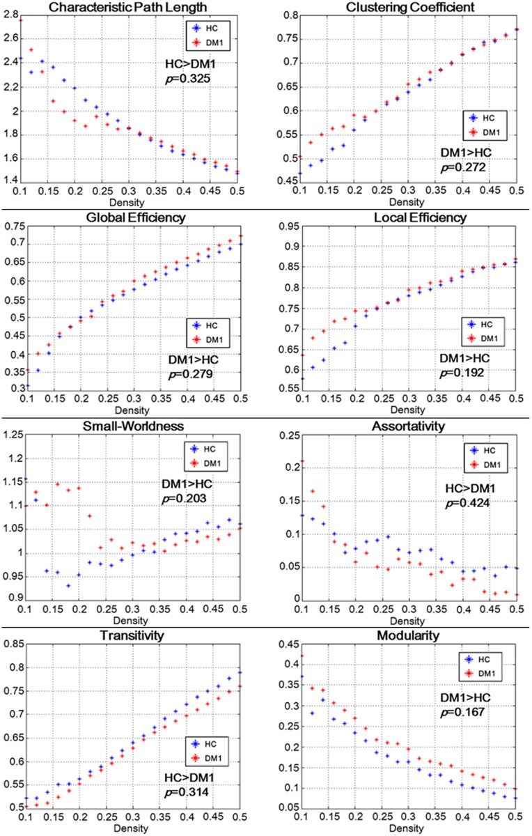
This study aimed to investigate abnormalities in structural covariance network constructed from gray matter volume in myotonic dystrophy type 1 (DM1) patients by using graph theoretical analysis for further clarification of the underlying mechanisms of central nervous system involvement. Twenty-eight DM1 patients (4 childhood onset, 10 juvenile onset, 14 adult onset), excluding three cases from 31 consecutive patients who underwent magnetic resonance imaging in a certain period, and 28 age- and sex- matched healthy control subjects were included in this study. The normalized gray matter images of both groups were subjected to voxel based morphometry (VBM) and Graph Analysis Toolbox for graph theoretical analysis. VBM revealed extensive gray matter atrophy in DM1 patients, including cortical and subcortical structures. On graph theoretical analysis, there were no significant differences between DM1 and control groups in terms of the global measures of connectivity. Betweenness centrality was increased in several regions including the left fusiform gyrus, whereas it was decreased in the right striatum. The absence of significant differences between the groups in global network measurements on graph theoretical analysis is consistent with the fact that the general cognitive function is preserved in DM1 patients. In DM1 patients, increased connectivity in the left fusiform gyrus and decreased connectivity in the right striatum might be associated with impairment in face perception and theory of mind, and schizotypal-paranoid personality traits, respectively.
本研究旨在通过使用图论分析来研究1型强直性肌营养不良(DM1)患者基于灰质体积构建的结构协方差网络异常,以进一步阐明中枢神经系统受累的潜在机制。本研究纳入了28例DM1患者(4例儿童期起病,10例青少年期起病,14例成年期起病),排除了某一时期连续接受磁共振成像检查的31例患者中的3例,以及28例年龄和性别匹配的健康对照者。对两组的标准化灰质图像进行基于体素的形态学测量(VBM)和用于图论分析的图形分析工具箱分析。VBM显示DM1患者存在广泛的灰质萎缩,包括皮质和皮质下结构。在图论分析中,DM1组和对照组在连接性的整体测量方面没有显著差异。包括左侧梭状回在内的几个区域的中间中心性增加,而右侧纹状体的中间中心性降低。图论分析中两组在整体网络测量上没有显著差异,这与DM1患者一般认知功能保留的事实一致。在DM1患者中,左侧梭状回连接性增加和右侧纹状体连接性降低可能分别与面部感知障碍、心理理论以及分裂样偏执型人格特质有关。