Institute of Molecular Bioimaging and Physiology, National Research Council (IBFM-CNR), Milan, Italy.
Medical Physics Unit, IRCCS Fondazione S. Maugeri, Pavia, Italy.
Contrast Media Mol Imaging. 2017 Sep 7;2017:3461684. doi: 10.1155/2017/3461684. eCollection 2017.
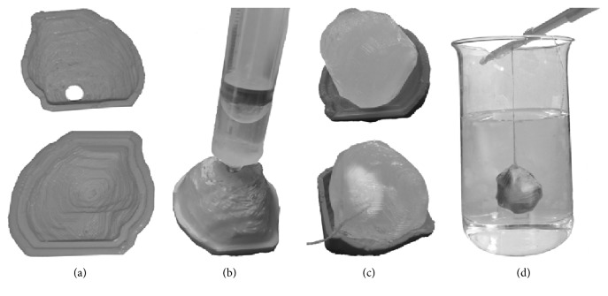
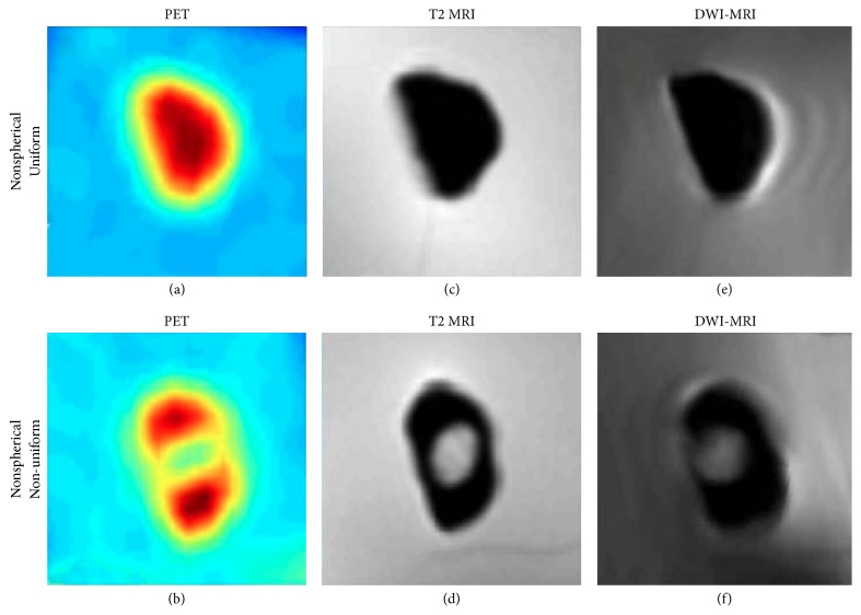
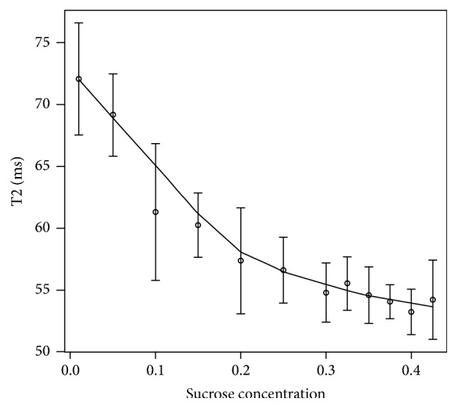
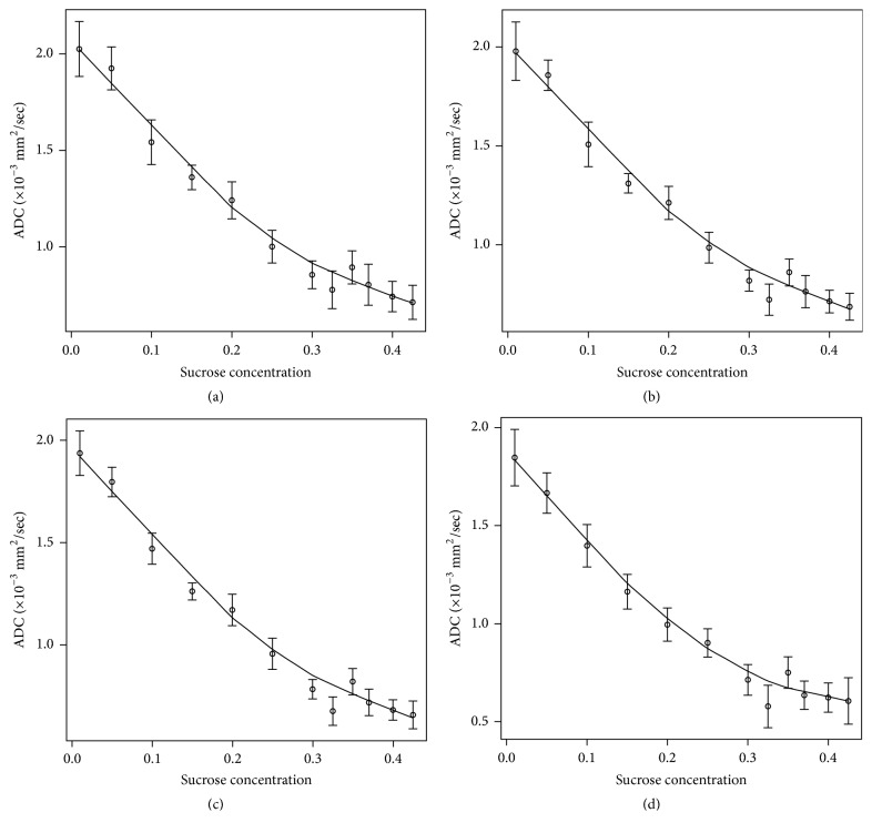
The aim of this work was to develop a method to manufacture oncological phantoms for quantitation purposes in 18F-FDG PET and DW-MRI studies. Radioactive and diffusion materials were prepared using a mixture of agarose and sucrose radioactive gels. T2 relaxation and diffusion properties of gels at different sucrose concentrations were evaluated. Realistic oncological lesions were created using 3D-printed plastic molds filled with the gel mixture. Once solidified, gels were extracted from molds and immersed in a low-radioactivity gel simulating normal background tissue. A breast cancer phantom was manufactured using the proposed method as an exploratory feasibility study, including several realistic oncological configurations in terms of both radioactivity and diffusion. The phantom was acquired in PET with 18F-FDG, immediately after solidification, and in DW-MRI the following day. Functional volumes characterizing the simulated BC lesions were segmented from PET and DW-MRI images. Measured radioactive uptake and ADC values were compared with gold standards. Phantom preparation was straightforward, and the time schedule was compatible with both PET and MRI measurements. Lesions appeared on 18F-FDG PET and DW-MRI images as expected, without visible artifacts. Lesion functional parameters revealed the phantom's potential for validating quantification methods, in particular for new generation hybrid PET-MRI systems.
这项工作的目的是开发一种方法,用于制造用于 18F-FDG PET 和 DW-MRI 研究定量目的的肿瘤体模。放射性和扩散材料是使用琼脂糖和蔗糖放射性凝胶混合物制备的。评估了不同蔗糖浓度下凝胶的 T2 弛豫和扩散特性。使用填充有凝胶混合物的 3D 打印塑料模具创建逼真的肿瘤病变。凝胶从模具中取出并浸入低放射性凝胶中,以模拟正常背景组织。使用提出的方法制造了乳腺癌体模,作为探索性可行性研究,包括放射性和扩散方面的几种现实肿瘤学配置。体模在凝固后立即用 18F-FDG 进行 PET 采集,并在第二天进行 DW-MRI 采集。从 PET 和 DW-MRI 图像中分割出模拟 BC 病变的功能体积。测量的放射性摄取和 ADC 值与金标准进行了比较。体模制备简单,时间安排与 PET 和 MRI 测量兼容。肿瘤病变在 18F-FDG PET 和 DW-MRI 图像上的出现与预期一致,没有可见伪影。病变功能参数显示了该体模验证定量方法的潜力,特别是对于新一代的 PET-MRI 混合系统。