• 文献检索
  • 文档翻译
  • 深度研究
  • 学术资讯
  • Suppr Zotero 插件Zotero 插件
  • 邀请有礼
  • 套餐&价格
  • 历史记录
应用&插件
Suppr Zotero 插件Zotero 插件浏览器插件Mac 客户端Windows 客户端微信小程序
定价
高级版会员购买积分包购买API积分包
服务
文献检索文档翻译深度研究API 文档MCP 服务
关于我们
关于 Suppr公司介绍联系我们用户协议隐私条款
关注我们

Suppr 超能文献

核心技术专利:CN118964589B侵权必究
粤ICP备2023148730 号-1Suppr @ 2026

文献检索

告别复杂PubMed语法,用中文像聊天一样搜索,搜遍4000万医学文献。AI智能推荐,让科研检索更轻松。

立即免费搜索

文件翻译

保留排版,准确专业,支持PDF/Word/PPT等文件格式,支持 12+语言互译。

免费翻译文档

深度研究

AI帮你快速写综述,25分钟生成高质量综述,智能提取关键信息,辅助科研写作。

立即免费体验

揭示 TPX2 刺激微管组装的结构基础

Structural insight into TPX2-stimulated microtubule assembly.

机构信息

Molecular Biophysics and Integrative Bioimaging Division, Lawrence Berkeley National Laboratory, Berkeley, United States.

The Francis Crick Institute, London, United Kingdom.

出版信息

Elife. 2017 Nov 9;6:e30959. doi: 10.7554/eLife.30959.

DOI:10.7554/eLife.30959
PMID:29120325
原文链接:https://pmc.ncbi.nlm.nih.gov/articles/PMC5679754/
Abstract

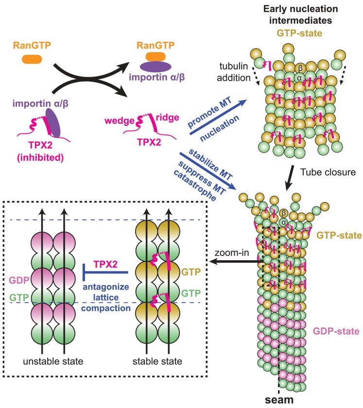
During mitosis and meiosis, microtubule (MT) assembly is locally upregulated by the chromatin-dependent Ran-GTP pathway. One of its key targets is the MT-associated spindle assembly factor TPX2. The molecular mechanism of how TPX2 stimulates MT assembly remains unknown because structural information about the interaction of TPX2 with MTs is lacking. Here, we determine the cryo-electron microscopy structure of a central region of TPX2 bound to the MT surface. TPX2 uses two flexibly linked elements ('ridge' and 'wedge') in a novel interaction mode to simultaneously bind across longitudinal and lateral tubulin interfaces. These MT-interacting elements overlap with the binding site of importins on TPX2. Fluorescence microscopy-based in vitro reconstitution assays reveal that this interaction mode is critical for MT binding and facilitates MT nucleation. Together, our results suggest a molecular mechanism of how the Ran-GTP gradient can regulate TPX2-dependent MT formation.

摘要

在有丝分裂和减数分裂过程中,微管(MT)的组装由染色质依赖的 Ran-GTP 途径局部上调。其关键靶标之一是与 MT 相关的纺锤体组装因子 TPX2。TPX2 如何刺激 MT 组装的分子机制尚不清楚,因为缺乏关于 TPX2 与 MT 相互作用的结构信息。在这里,我们确定了与 MT 表面结合的 TPX2 中心区域的冷冻电子显微镜结构。TPX2 使用两个灵活连接的元件(“脊”和“楔”)以新颖的相互作用模式同时结合在纵向和横向微管界面上。这些与 TPX2 上进口蛋白结合位点重叠的 MT 相互作用元件。基于荧光显微镜的体外重建测定表明,这种相互作用模式对于 MT 结合至关重要,并促进 MT 成核。总之,我们的结果表明了 Ran-GTP 梯度如何调节 TPX2 依赖性 MT 形成的分子机制。

https://cdn.ncbi.nlm.nih.gov/pmc/blobs/2a62/5679754/64ead7b1153a/elife-30959-fig6-figsupp1.jpg
https://cdn.ncbi.nlm.nih.gov/pmc/blobs/2a62/5679754/ae0439dd3561/elife-30959-fig1.jpg
https://cdn.ncbi.nlm.nih.gov/pmc/blobs/2a62/5679754/b7165b747283/elife-30959-fig1-figsupp1.jpg
https://cdn.ncbi.nlm.nih.gov/pmc/blobs/2a62/5679754/b755ee73d14d/elife-30959-fig1-figsupp2.jpg
https://cdn.ncbi.nlm.nih.gov/pmc/blobs/2a62/5679754/77e450af2413/elife-30959-fig1-figsupp3.jpg
https://cdn.ncbi.nlm.nih.gov/pmc/blobs/2a62/5679754/d29ddc5c982b/elife-30959-fig1-figsupp4.jpg
https://cdn.ncbi.nlm.nih.gov/pmc/blobs/2a62/5679754/7fc0ae8c18a8/elife-30959-fig1-figsupp5.jpg
https://cdn.ncbi.nlm.nih.gov/pmc/blobs/2a62/5679754/0554bb7b2b89/elife-30959-fig2.jpg
https://cdn.ncbi.nlm.nih.gov/pmc/blobs/2a62/5679754/ab3332f49070/elife-30959-fig3.jpg
https://cdn.ncbi.nlm.nih.gov/pmc/blobs/2a62/5679754/8cea710f71f1/elife-30959-fig3-figsupp1.jpg
https://cdn.ncbi.nlm.nih.gov/pmc/blobs/2a62/5679754/d7fe61ccd42b/elife-30959-fig4.jpg
https://cdn.ncbi.nlm.nih.gov/pmc/blobs/2a62/5679754/69cb55bfc39f/elife-30959-fig5.jpg
https://cdn.ncbi.nlm.nih.gov/pmc/blobs/2a62/5679754/7af94e6c62ab/elife-30959-fig5-figsupp1.jpg
https://cdn.ncbi.nlm.nih.gov/pmc/blobs/2a62/5679754/634f53cdb821/elife-30959-fig5-figsupp2.jpg
https://cdn.ncbi.nlm.nih.gov/pmc/blobs/2a62/5679754/5d01daad6972/elife-30959-fig6.jpg
https://cdn.ncbi.nlm.nih.gov/pmc/blobs/2a62/5679754/64ead7b1153a/elife-30959-fig6-figsupp1.jpg

相似文献

1
Structural insight into TPX2-stimulated microtubule assembly.揭示 TPX2 刺激微管组装的结构基础
Elife. 2017 Nov 9;6:e30959. doi: 10.7554/eLife.30959.
2
Characterization of the TPX2 domains involved in microtubule nucleation and spindle assembly in Xenopus egg extracts.非洲爪蟾卵提取物中参与微管成核和纺锤体组装的TPX2结构域的特性分析
Mol Biol Cell. 2004 Dec;15(12):5318-28. doi: 10.1091/mbc.e04-05-0385. Epub 2004 Sep 22.
3
Microtubule nucleation in mitosis by a RanGTP-dependent protein complex.有丝分裂中由RanGTP依赖性蛋白复合物介导的微管成核作用。
Curr Biol. 2015 Jan 19;25(2):131-140. doi: 10.1016/j.cub.2014.11.025. Epub 2014 Dec 18.
4
Diverse microtubule-binding repeats regulate TPX2 activities at distinct locations within the spindle.多种微管结合重复序列在纺锤体内不同位置调节TPX2活性。
J Cell Biol. 2025 Mar 3;224(3). doi: 10.1083/jcb.202404025. Epub 2025 Jan 16.
5
Structural analysis of the role of TPX2 in branching microtubule nucleation.TPX2在分支微管成核作用中的结构分析
J Cell Biol. 2017 Apr 3;216(4):983-997. doi: 10.1083/jcb.201607060. Epub 2017 Mar 6.
6
Identification of a TPX2-like microtubule-associated protein in Drosophila.在果蝇中鉴定出一个与微管相关的 TPX2 样蛋白。
PLoS One. 2011;6(11):e28120. doi: 10.1371/journal.pone.0028120. Epub 2011 Nov 30.
7
Complementary activities of TPX2 and chTOG constitute an efficient importin-regulated microtubule nucleation module.TPX2和chTOG的互补活性构成了一个有效的输入蛋白调节的微管成核模块。
Nat Cell Biol. 2015 Nov;17(11):1422-34. doi: 10.1038/ncb3241. Epub 2015 Sep 28.
8
Interaction of spindle assembly factor TPX2 with importins-α/β inhibits protein phase separation.纺锤体组装因子 TPX2 与 importins-α/β 的相互作用抑制了蛋白质相分离。
J Biol Chem. 2021 Sep;297(3):100998. doi: 10.1016/j.jbc.2021.100998. Epub 2021 Jul 21.
9
Ran-dependent TPX2 activation promotes acentrosomal microtubule nucleation in neurons.Ran 依赖性的 TPX2 激活促进神经元中无中心体微管的核形成。
Sci Rep. 2017 Feb 13;7:42297. doi: 10.1038/srep42297.
10
Ran modulates spindle assembly by regulating a subset of TPX2 and Kid activities including Aurora A activation.Ran通过调节TPX2和Kid活性的一个子集(包括极光激酶A激活)来调节纺锤体组装。
J Cell Sci. 2003 Dec 1;116(Pt 23):4791-8. doi: 10.1242/jcs.00798.

引用本文的文献

1
Conserved function of the HAUS6 calponin homology domain in anchoring augmin for microtubule branching.HAUS6的钙调蛋白同源结构域在锚定augmin以促进微管分支中的保守功能。
Nat Commun. 2025 Aug 22;16(1):7845. doi: 10.1038/s41467-025-63165-z.
2
CFAP100 couples microtubule glutamylation to spindle pole integrity in keratinocytes to promote epidermal development.CFAP100将微管蛋白谷氨酰胺化与角质形成细胞中的纺锤体极完整性联系起来,以促进表皮发育。
Nat Commun. 2025 Jul 1;16(1):5591. doi: 10.1038/s41467-025-60677-6.
3
Meiotic divisions and round spermatid formation do not require centriole duplication in mice.

本文引用的文献

1
The Centrosome Is a Selective Condensate that Nucleates Microtubules by Concentrating Tubulin.中心体是一种选择性凝聚物,通过浓缩微管蛋白来成核微管。
Cell. 2017 Jun 1;169(6):1066-1077.e10. doi: 10.1016/j.cell.2017.05.028.
2
Structural analysis of the role of TPX2 in branching microtubule nucleation.TPX2在分支微管成核作用中的结构分析
J Cell Biol. 2017 Apr 3;216(4):983-997. doi: 10.1083/jcb.201607060. Epub 2017 Mar 6.
3
The C-terminal domain of TPX2 is made of alpha-helical tandem repeats.TPX2的C末端结构域由α螺旋串联重复序列组成。
减数分裂和圆形精子细胞的形成在小鼠中不需要中心粒复制。
PLoS Genet. 2025 Apr 28;21(4):e1011698. doi: 10.1371/journal.pgen.1011698. eCollection 2025 Apr.
4
An Aurora kinase A-BOD1L1-PP2A B56 axis promotes chromosome segregation fidelity.极光激酶A-BOD1L1-蛋白磷酸酶2A B56轴促进染色体分离保真度。
Cell Rep. 2025 Feb 25;44(2):115317. doi: 10.1016/j.celrep.2025.115317. Epub 2025 Feb 18.
5
NKX6.3 modulation of mitotic dynamics and genomic stability in gastric carcinogenesis.NKX6.3对胃癌发生过程中细胞有丝分裂动力学和基因组稳定性的调控
Cell Commun Signal. 2025 Jan 20;23(1):35. doi: 10.1186/s12964-025-02030-4.
6
Diverse microtubule-binding repeats regulate TPX2 activities at distinct locations within the spindle.多种微管结合重复序列在纺锤体内不同位置调节TPX2活性。
J Cell Biol. 2025 Mar 3;224(3). doi: 10.1083/jcb.202404025. Epub 2025 Jan 16.
7
Maternal ELL3 loss-of-function leads to oocyte aneuploidy and early miscarriage.母体ELL3功能丧失导致卵母细胞非整倍体和早期流产。
Nat Struct Mol Biol. 2025 Feb;32(2):381-392. doi: 10.1038/s41594-024-01471-8. Epub 2025 Jan 16.
8
Phase separation of microtubule-binding proteins - implications for neuronal function and disease.微管结合蛋白的相分离——对神经元功能和疾病的影响
J Cell Sci. 2024 Dec 15;137(24). doi: 10.1242/jcs.263470. Epub 2024 Dec 13.
9
Microtubule choreography: spindle self-organization during cell division.微管编排:细胞分裂过程中的纺锤体自我组织
Biophys Rev. 2024 Sep 30;16(5):613-624. doi: 10.1007/s12551-024-01236-z. eCollection 2024 Oct.
10
HURP facilitates spindle assembly by stabilizing microtubules and working synergistically with TPX2.HURP 通过稳定微管并与 TPX2 协同作用来促进纺锤体的组装。
Nat Commun. 2024 Nov 8;15(1):9689. doi: 10.1038/s41467-024-53630-6.
BMC Struct Biol. 2016 Oct 26;16(1):17. doi: 10.1186/s12900-016-0070-8.
4
EB1 interacts with outwardly curved and straight regions of the microtubule lattice.EB1 与微管晶格的外凸弯曲区和直线区相互作用。
Nat Cell Biol. 2016 Oct;18(10):1102-8. doi: 10.1038/ncb3412. Epub 2016 Sep 12.
5
Near-atomic cryo-EM structure of PRC1 bound to the microtubule.与微管结合的PRC1的近原子分辨率冷冻电镜结构
Proc Natl Acad Sci U S A. 2016 Aug 23;113(34):9430-9. doi: 10.1073/pnas.1609903113. Epub 2016 Aug 4.
6
Suppression of microtubule assembly kinetics by the mitotic protein TPX2.有丝分裂蛋白TPX2对微管组装动力学的抑制作用。
J Cell Sci. 2016 Apr 1;129(7):1319-28. doi: 10.1242/jcs.178806. Epub 2016 Feb 11.
7
Visualizing microtubule structural transitions and interactions with associated proteins.可视化微管结构转变及其与相关蛋白的相互作用。
Curr Opin Struct Biol. 2016 Apr;37:90-6. doi: 10.1016/j.sbi.2015.12.009. Epub 2016 Jan 21.
8
The RanGTP Pathway: From Nucleo-Cytoplasmic Transport to Spindle Assembly and Beyond.RanGTP 通路:从核质转运到纺锤体组装及其他。
Front Cell Dev Biol. 2016 Jan 11;3:82. doi: 10.3389/fcell.2015.00082. eCollection 2015.
9
A new protocol to accurately determine microtubule lattice seam location.一种准确确定微管晶格接缝位置的新方案。
J Struct Biol. 2015 Nov;192(2):245-54. doi: 10.1016/j.jsb.2015.09.015. Epub 2015 Sep 28.
10
Complementary activities of TPX2 and chTOG constitute an efficient importin-regulated microtubule nucleation module.TPX2和chTOG的互补活性构成了一个有效的输入蛋白调节的微管成核模块。
Nat Cell Biol. 2015 Nov;17(11):1422-34. doi: 10.1038/ncb3241. Epub 2015 Sep 28.