Andersen S H, Vervelde L, Sutton K, Norup L R, Wattrang E, Juul-Madsen H R, Dalgaard T S
Department of Animal Science, Aarhus University, Tjele, Denmark.
The Roslin Institute and Royal (Dick) School of Veterinary Sciences, University of Edinburgh, Easter Bush, Midlothian, UK.
Vet Immunol Immunopathol. 2017 Dec;193-194:18-28. doi: 10.1016/j.vetimm.2017.10.001. Epub 2017 Oct 10.
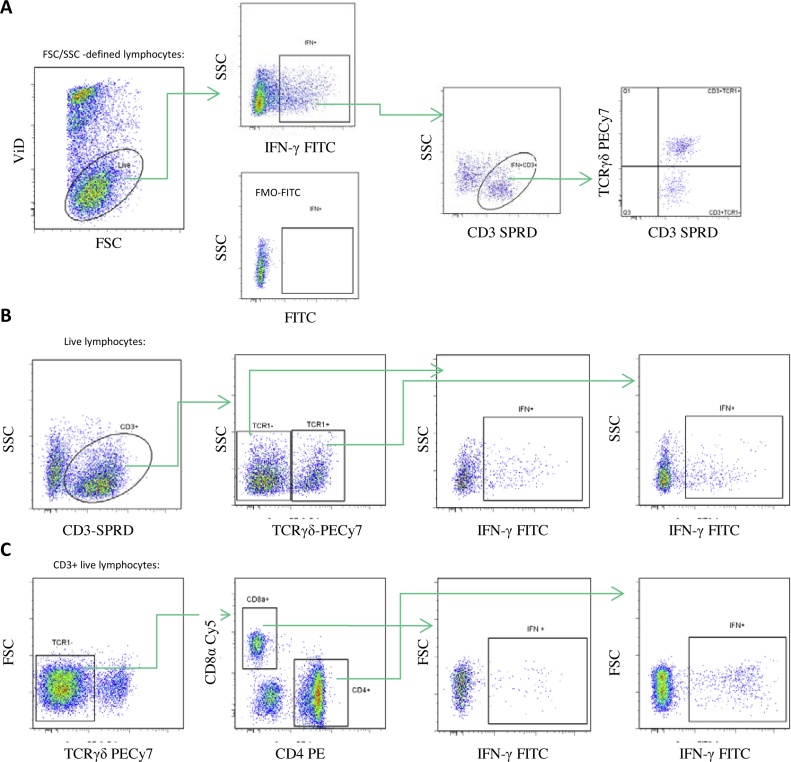
The aim of this study was to optimise and evaluate an intracellular cytokine staining (ICS) assay for assessment of T cell IFN-γ responses in chickens vaccinated against Newcastle disease (ND). We aimed to validate currently available antibodies to chicken IFN-γ using transfected CHO cells. Moreover, this ICS assay was evaluated for use to detect mitogen and antigen induced IFN-γ production in chicken peripheral blood leucocytes. Chickens from an inbred white leghorn line containing two MHC haplotypes, B19 and B21, were divided into three experimental groups; one group was kept as naive controls, one group was vaccinated intramuscularly twice with a commercial inactivated ND virus (NDV) vaccine, and the last group was vaccinated orally twice with a commercial live attenuated NDV vaccine. PBMC were ex vivo stimulated with ConA or with NDV antigen. The ICS assay was used to determine the phenotype and frequency of IFN-γ positive cells. ConA stimulation induced extensive IFN-γ production in both CD3TCRγδ (γδ T cells) cells and CD3TCRγδ cells (αβ T cells), but no significant differences were observed between the experimental groups. Furthermore, a large proportion of the IFN-γ producing cells were CD3 indicating that other cells than classic T cells, secreted this cytokine. NDV antigen stimulation induced IFN-γ production but to a lower extent than ConA and with a large variation between individuals. The CD3TCR1γδCD8α (CTL) population produced the highest NDV specific IFN-γ responses, with significantly elevated levels of IFN-γ producing cells in the B19 chickens vaccinated orally with live attenuated NDV vaccine. This was not the case in the B21 animals, indicating a haplotype restricted variation. In contrast, the CD3TCR1γδCD4 (Th) population did not show a significant increase in IFN-γ production in NDV stimulated samples which was in part due to a high number of IFN-γ producing cells after incubation with medium alone. In conclusion, an ICS assay for phenotyping of IFN-γ producing chicken leukocytes was set up that proved useful in identifying cytokine producing cells upon either mitogen or antigen-specific stimulation.
本研究的目的是优化和评估一种细胞内细胞因子染色(ICS)检测方法,用于评估接种新城疫(ND)疫苗的鸡的T细胞IFN-γ反应。我们旨在使用转染的CHO细胞验证目前可用的抗鸡IFN-γ抗体。此外,对该ICS检测方法进行了评估,以检测鸡外周血白细胞中丝裂原和抗原诱导的IFN-γ产生。来自含有两种MHC单倍型B19和B21的近交白来航品系的鸡被分为三个实验组;一组作为未免疫对照,一组肌肉注射两次商业灭活ND病毒(NDV)疫苗,最后一组口服两次商业减毒活NDV疫苗。外周血单核细胞(PBMC)在体外分别用刀豆蛋白A(ConA)或NDV抗原刺激。ICS检测方法用于确定IFN-γ阳性细胞的表型和频率。ConA刺激在CD3TCRγδ(γδ T细胞)细胞和CD3TCRγδ细胞(αβ T细胞)中均诱导了大量的IFN-γ产生,但实验组之间未观察到显著差异。此外,大部分产生IFN-γ的细胞为CD3阳性,这表明除经典T细胞外的其他细胞也分泌这种细胞因子。NDV抗原刺激诱导了IFN-γ产生,但程度低于ConA,且个体间差异较大。CD3TCR1γδCD8α(CTL)群体产生的NDV特异性IFN-γ反应最高,口服减毒活NDV疫苗的B19鸡中产生IFN-γ的细胞水平显著升高。B21动物中情况并非如此,表明存在单倍型限制的差异。相比之下,CD3TCR1γδCD4(Th)群体在NDV刺激的样本中IFN-γ产生没有显著增加,部分原因是仅用培养基孵育后就有大量产生IFN-γ的细胞。总之,建立了一种用于对产生IFN-γ的鸡白细胞进行表型分析的ICS检测方法,该方法被证明在识别丝裂原或抗原特异性刺激后产生细胞因子的细胞方面很有用。