• 文献检索
  • 文档翻译
  • 深度研究
  • 学术资讯
  • Suppr Zotero 插件Zotero 插件
  • 邀请有礼
  • 套餐&价格
  • 历史记录
应用&插件
Suppr Zotero 插件Zotero 插件浏览器插件Mac 客户端Windows 客户端微信小程序
定价
高级版会员购买积分包购买API积分包
服务
文献检索文档翻译深度研究API 文档MCP 服务
关于我们
关于 Suppr公司介绍联系我们用户协议隐私条款
关注我们

Suppr 超能文献

核心技术专利:CN118964589B侵权必究
粤ICP备2023148730 号-1Suppr @ 2026

文献检索

告别复杂PubMed语法,用中文像聊天一样搜索,搜遍4000万医学文献。AI智能推荐,让科研检索更轻松。

立即免费搜索

文件翻译

保留排版,准确专业,支持PDF/Word/PPT等文件格式,支持 12+语言互译。

免费翻译文档

深度研究

AI帮你快速写综述,25分钟生成高质量综述,智能提取关键信息,辅助科研写作。

立即免费体验

染色体分离发生在卵母细胞中微管的推动下。

Chromosome segregation occurs by microtubule pushing in oocytes.

机构信息

Institut Jacques Monod, CNRS, UMR 7592, University Paris Diderot, Sorbonne Paris Cité, 75205, Paris, France.

Department of Pathology and Cell Biology, Columbia University Medical Center, New York, NY, 10032, USA.

出版信息

Nat Commun. 2017 Nov 14;8(1):1499. doi: 10.1038/s41467-017-01539-8.

DOI:10.1038/s41467-017-01539-8
PMID:29133801
原文链接:https://pmc.ncbi.nlm.nih.gov/articles/PMC5684144/
Abstract

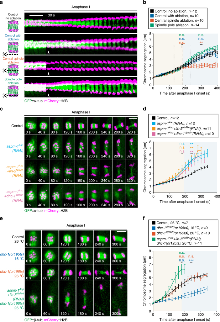
During cell division, spindle microtubules ensure an equal repartition of chromosomes between the two daughter cells. While the kinetochore-dependent mechanisms that drive mitotic chromosome segregation are well understood, in oocytes of most species atypical spindles assembled in absence of centrosomes entail poorly understood mechanisms of chromosome segregation. In particular, the structure(s) responsible for force generation during meiotic chromosome separation in oocytes is unclear. Using quantitative light microscopy, electron tomography, laser-mediated ablation, and genetic perturbations in the Caenorhabditis elegans oocyte, we studied the mechanism of chromosome segregation in meiosis. We find spindle poles are largely dispensable, and in fact act as brakes for chromosome segregation. Instead, our results suggest that CLS-2-dependent microtubules of the meiotic central spindle, located between the segregating chromosomes and aligned along the axis of segregation, are essential. Our results support a model in which inter-chromosomal microtubules of the central spindle push chromosomes apart during meiotic anaphase in oocytes.

摘要

在细胞分裂过程中,纺锤体微管确保染色体在两个子细胞之间均等分配。虽然驱动有丝分裂染色体分离的着丝粒依赖性机制已被很好地理解,但在大多数物种的卵母细胞中,在没有中心体的情况下组装的非典型纺锤体涉及到染色体分离的机制尚不清楚。特别是,在卵母细胞中进行减数分裂染色体分离过程中产生力的结构尚不清楚。使用定量光显微镜、电子断层扫描、激光介导的消融以及在秀丽隐杆线虫卵母细胞中的遗传干扰,我们研究了减数分裂中的染色体分离机制。我们发现纺锤体极大部分是可有可无的,实际上它们充当了染色体分离的制动器。相反,我们的结果表明,位于分离染色体之间并沿着分离轴排列的减数分裂中心纺锤体上的 CLS-2 依赖性微管是必不可少的。我们的结果支持这样一种模型,即中心纺锤体的染色体间微管在卵母细胞的减数分裂后期将染色体推开。

https://cdn.ncbi.nlm.nih.gov/pmc/blobs/b674/5684144/2e79762d4bf4/41467_2017_1539_Fig5_HTML.jpg
https://cdn.ncbi.nlm.nih.gov/pmc/blobs/b674/5684144/4afdd4de9df4/41467_2017_1539_Fig1_HTML.jpg
https://cdn.ncbi.nlm.nih.gov/pmc/blobs/b674/5684144/867ccae5997c/41467_2017_1539_Fig2_HTML.jpg
https://cdn.ncbi.nlm.nih.gov/pmc/blobs/b674/5684144/a7f6e5e70972/41467_2017_1539_Fig3_HTML.jpg
https://cdn.ncbi.nlm.nih.gov/pmc/blobs/b674/5684144/373074c15937/41467_2017_1539_Fig4_HTML.jpg
https://cdn.ncbi.nlm.nih.gov/pmc/blobs/b674/5684144/2e79762d4bf4/41467_2017_1539_Fig5_HTML.jpg

相似文献

1
Chromosome segregation occurs by microtubule pushing in oocytes.染色体分离发生在卵母细胞中微管的推动下。
Nat Commun. 2017 Nov 14;8(1):1499. doi: 10.1038/s41467-017-01539-8.
2
A Switch in Microtubule Orientation during C. elegans Meiosis.线虫减数分裂过程中微管取向的转换。
Curr Biol. 2018 Sep 24;28(18):2991-2997.e2. doi: 10.1016/j.cub.2018.07.012. Epub 2018 Sep 6.
3
Multiple motors cooperate to establish and maintain acentrosomal spindle bipolarity in oocyte meiosis.多个马达协同作用以建立和维持卵母细胞减数分裂中的无中心体纺锤体双极性。
Elife. 2022 Feb 11;11:e72872. doi: 10.7554/eLife.72872.
4
KLP-7 acts through the Ndc80 complex to limit pole number in C. elegans oocyte meiotic spindle assembly.KLP-7通过Ndc80复合体发挥作用,以限制秀丽隐杆线虫卵母细胞减数分裂纺锤体组装中的极数。
J Cell Biol. 2015 Sep 14;210(6):917-32. doi: 10.1083/jcb.201412010.
5
Central-spindle microtubules are strongly coupled to chromosomes during both anaphase A and anaphase B.有丝分裂后期 A 和有丝分裂后期 B 期间,中心纺锤体微管与染色体紧密结合。
Mol Biol Cell. 2019 Sep 1;30(19):2503-2514. doi: 10.1091/mbc.E19-01-0074. Epub 2019 Jul 24.
6
A kinetochore-independent mechanism drives anaphase chromosome separation during acentrosomal meiosis.无着丝粒的机制驱动非中心体减数分裂后期染色体的分离。
Nat Cell Biol. 2010 Sep;12(9):894-901. doi: 10.1038/ncb2093. Epub 2010 Aug 22.
7
Synergistic stabilization of microtubules by BUB-1, HCP-1, and CLS-2 controls microtubule pausing and meiotic spindle assembly.BUB-1、HCP-1 和 CLS-2 通过协同稳定微管控制微管停顿和减数分裂纺锤体的组装。
Elife. 2023 Feb 17;12:e82579. doi: 10.7554/eLife.82579.
8
Sumoylation regulates protein dynamics during meiotic chromosome segregation in oocytes.SUMOylation 在卵母细胞减数分裂染色体分离过程中调节蛋白质动力学。
J Cell Sci. 2019 Jul 18;132(14):jcs232330. doi: 10.1242/jcs.232330.
9
BUB-1 promotes amphitelic chromosome biorientation via multiple activities at the kinetochore.BUB-1 通过在动粒上的多种活性促进联会染色体的双定向。
Elife. 2018 Dec 14;7:e40690. doi: 10.7554/eLife.40690.
10
Kinetochore component function in C. elegans oocytes revealed by 4D tracking of holocentric chromosomes.利用整体染色体的 4D 追踪揭示线虫卵母细胞中的着丝粒组件功能。
Nat Commun. 2023 Jul 7;14(1):4032. doi: 10.1038/s41467-023-39702-z.

引用本文的文献

1
Discovery of unique mitotic mechanisms in .在……中发现独特的有丝分裂机制。 (你提供的原文不完整,“in”后面缺少具体内容)
Open Biol. 2025 Aug;15(8):250096. doi: 10.1098/rsob.250096. Epub 2025 Aug 6.
2
Inward transport of organelles drives outward migration of the spindle during C. elegans meiosis.在秀丽隐杆线虫减数分裂过程中,细胞器的内向运输驱动纺锤体向外迁移。
Cell Rep. 2025 Apr 22;44(4):115458. doi: 10.1016/j.celrep.2025.115458. Epub 2025 Mar 22.
3
An interkinetic envelope surrounds chromosomes between meiosis I and II in C. elegans oocytes.

本文引用的文献

1
Molecular mechanism of dynein recruitment to kinetochores by the Rod-Zw10-Zwilch complex and Spindly.动力蛋白通过Rod-Zw10-Zwilch复合体和Spindly被募集到动粒的分子机制。
J Cell Biol. 2017 Apr 3;216(4):943-960. doi: 10.1083/jcb.201610108. Epub 2017 Mar 20.
2
A Molecular View of Kinetochore Assembly and Function.动粒组装与功能的分子视角
Biology (Basel). 2017 Jan 24;6(1):5. doi: 10.3390/biology6010005.
3
Assembly of Caenorhabditis elegans acentrosomal spindles occurs without evident microtubule-organizing centers and requires microtubule sorting by KLP-18/kinesin-12 and MESP-1.
在秀丽隐杆线虫卵母细胞中,减数分裂I和II之间,一种运动间期包膜围绕着染色体。
J Cell Biol. 2025 Mar 3;224(3). doi: 10.1083/jcb.202403125. Epub 2024 Dec 26.
4
An interkinetic envelope surrounds chromosomes between meiosis I and II in oocytes.在卵母细胞中,减数分裂I和II之间,一个运动间期包膜围绕着染色体。
bioRxiv. 2024 Oct 21:2024.10.19.619195. doi: 10.1101/2024.10.19.619195.
5
Unveiling inter-embryo variability in spindle length over time: Towards quantitative phenotype analysis.揭示胚胎间纺锤体长度随时间的变化:迈向定量表型分析。
PLoS Comput Biol. 2024 Sep 5;20(9):e1012330. doi: 10.1371/journal.pcbi.1012330. eCollection 2024 Sep.
6
AlphaFold2-guided engineering of split-GFP technology enables labeling of endogenous tubulins across species while preserving function.基于 AlphaFold2 的 split-GFP 技术工程改造实现了跨物种标记内源性微管蛋白而不影响其功能。
PLoS Biol. 2024 Aug 19;22(8):e3002615. doi: 10.1371/journal.pbio.3002615. eCollection 2024 Aug.
7
Raman Spectroscopy Reveals Photobiomodulation-Induced α-Helix to β-Sheet Transition in Tubulins: Potential Implications for Alzheimer's and Other Neurodegenerative Diseases.拉曼光谱揭示光生物调节诱导微管蛋白从α-螺旋向β-折叠转变:对阿尔茨海默病和其他神经退行性疾病的潜在影响。
Nanomaterials (Basel). 2024 Jun 26;14(13):1093. doi: 10.3390/nano14131093.
8
RNF20 Regulates Oocyte Meiotic Spindle Assembly by Recruiting TPM3 to Centromeres and Spindle Poles.RNF20 通过招募 TPM3 到着丝粒和纺锤体两极来调节卵母细胞减数分裂纺锤体的组装。
Adv Sci (Weinh). 2024 Apr;11(13):e2306986. doi: 10.1002/advs.202306986. Epub 2024 Jan 19.
9
F-actin coordinates spindle morphology and function in Drosophila meiosis.F-肌动蛋白协调果蝇减数分裂中纺锤体的形态和功能。
PLoS Genet. 2024 Jan 11;20(1):e1011111. doi: 10.1371/journal.pgen.1011111. eCollection 2024 Jan.
10
Redundant microtubule crosslinkers prevent meiotic spindle bending to ensure diploid offspring in C. elegans.冗余的微管交联蛋白防止减数分裂纺锤体弯曲,以确保线虫产生二倍体后代。
PLoS Genet. 2023 Dec 27;19(12):e1011090. doi: 10.1371/journal.pgen.1011090. eCollection 2023 Dec.
秀丽隐杆线虫无中心体纺锤体的组装在没有明显微管组织中心的情况下发生,并且需要通过KLP-18/驱动蛋白-12和MESP-1进行微管分选。
Mol Biol Cell. 2016 Oct 15;27(20):3122-3131. doi: 10.1091/mbc.E16-05-0291. Epub 2016 Aug 24.
4
A novel chromosome segregation mechanism during female meiosis.雌性减数分裂过程中的一种新型染色体分离机制。
Mol Biol Cell. 2016 Aug 15;27(16):2576-89. doi: 10.1091/mbc.E16-05-0331. Epub 2016 Jun 22.
5
Microcephaly protein Asp focuses the minus ends of spindle microtubules at the pole and within the spindle.小头蛋白Asp将纺锤体微管的负端聚集在纺锤体两极及纺锤体内。
J Cell Biol. 2015 Dec 7;211(5):999-1009. doi: 10.1083/jcb.201507001.
6
Kinetochore-independent chromosome segregation driven by lateral microtubule bundles.由横向微管束驱动的不依赖动粒的染色体分离
Elife. 2015 May 30;4:e06462. doi: 10.7554/eLife.06462.
7
Kinetochore components are required for central spindle assembly.着丝粒组件是中心纺锤体组装所必需的。
Nat Cell Biol. 2015 May;17(5):697-705. doi: 10.1038/ncb3150. Epub 2015 Apr 13.
8
Chromatids segregate without centrosomes during Caenorhabditis elegans mitosis in a Ran- and CLASP-dependent manner.在秀丽隐杆线虫有丝分裂过程中,染色单体在没有中心体的情况下以依赖于Ran和CLASP的方式进行分离。
Mol Biol Cell. 2015 Jun 1;26(11):2020-9. doi: 10.1091/mbc.E14-12-1577. Epub 2015 Apr 1.
9
Automated stitching of microtubule centerlines across serial electron tomograms.跨序列电子断层扫描的微管中心线自动拼接
PLoS One. 2014 Dec 1;9(12):e113222. doi: 10.1371/journal.pone.0113222. eCollection 2014.
10
Caenorhabditis elegans oocyte meiotic spindle pole assembly requires microtubule severing and the calponin homology domain protein ASPM-1.秀丽隐杆线虫卵母细胞减数分裂纺锤体极体组装需要微管切割和钙调蛋白同源结构域蛋白 ASPM-1。
Mol Biol Cell. 2014 Apr;25(8):1298-311. doi: 10.1091/mbc.E13-11-0687. Epub 2014 Feb 19.