Cho Hyung Joon, Kim Tae Kyun, Kang Seung-Baik, Do Min Uk, Chang Chong Bum
Department of Orthopaedic Surgery, Pusan National University Yangsan Hospital, 20 Geumo-ro, Mulgeum-eup, Gyeongsangnam-do, Yangsan-si, 50612, South Korea.
Joint Reconstruction Center, Seoul National University Bundang Hospital, 82, Gumi-ro 173beon-gil, Bundang-gu, Seongnam-si, Gyeonggi-do, 13620, South Korea.
BMC Musculoskelet Disord. 2017 Nov 14;18(1):448. doi: 10.1186/s12891-017-1822-8.
This cadaveric study aimed to demonstrate variation of the anterior cruciate ligament (ACL) tibial attachment in the sagittal plane, and to analyze the radiographic landmarks which predict the sagittal location of the ACL tibial attachment.
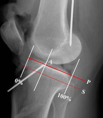
In 20 cadaveric knees, native ACLs were removed and the centers of the ACL tibial and femoral attachments were marked with metal pins. Full extension lateral radiographs were then obtained in each cadaveric knee. Using the full extension lateral radiographs, the sagittal location of the ACL tibial footprint center was estimated as a percentage in the Amis and Jakob's line. Several radiographic landmarks including the geometry of Blumensaat's line and the apex of the tibial eminence were measured. Then, the relationship between the variation of the sagittal location of the ACL tibial footprint and several radiographic landmarks were analyzed using Pearson's correlation analysis.
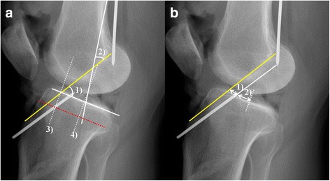
The average sagittal position of the native ACL tibial footprint was 40.9% (range: 38.0-45.0%). The line connecting the centers of the ACL footprint was nearly parallel to Blumensaat's line, with an average angle of 1.7° (range: 0-4.1°). In addition, the distance from the point where Blumensaat's line meets the tibial articular surface to the center of the ACL tibial footprint was almost consistent, at 7.6 mm on average (range: 6.4-8.7 mm). The correlation analysis revealed that the geometry of Blumensaat's line was significantly correlated with the sagittal location of the ACL tibial footprint.
The radiographic landmark that showed a significant correlation with the ACL tibial footprint in the full extension lateral radiographs was Blumensaat's line.
本尸体研究旨在展示前交叉韧带(ACL)胫骨附着点在矢状面的变异情况,并分析预测ACL胫骨附着点矢状位置的影像学标志。
在20具尸体膝关节中,移除天然ACL,并使用金属针标记ACL胫骨和股骨附着点的中心。然后对每个尸体膝关节进行完全伸展位的侧位X线片拍摄。利用完全伸展位侧位X线片,将ACL胫骨足迹中心的矢状位置估计为在阿米斯和雅各布线中的百分比。测量了包括布卢门萨特线的几何形状和胫骨髁间隆起顶点在内的几个影像学标志。然后,使用Pearson相关分析分析ACL胫骨足迹矢状位置的变异与几个影像学标志之间的关系。
天然ACL胫骨足迹的平均矢状位置为40.9%(范围:38.0 - 45.0%)。连接ACL足迹中心的线几乎与布卢门萨特线平行,平均角度为1.7°(范围:0 - 4.1°)。此外,布卢门萨特线与胫骨关节面相交点到ACL胫骨足迹中心的距离几乎一致,平均为7.6毫米(范围:6.4 - 8.7毫米)。相关分析显示,布卢门萨特线的几何形状与ACL胫骨足迹的矢状位置显著相关。
在完全伸展位侧位X线片中,与ACL胫骨足迹显著相关的影像学标志是布卢门萨特线。