• 文献检索
  • 文档翻译
  • 深度研究
  • 学术资讯
  • Suppr Zotero 插件Zotero 插件
  • 邀请有礼
  • 套餐&价格
  • 历史记录
应用&插件
Suppr Zotero 插件Zotero 插件浏览器插件Mac 客户端Windows 客户端微信小程序
定价
高级版会员购买积分包购买API积分包
服务
文献检索文档翻译深度研究API 文档MCP 服务
关于我们
关于 Suppr公司介绍联系我们用户协议隐私条款
关注我们

Suppr 超能文献

核心技术专利:CN118964589B侵权必究
粤ICP备2023148730 号-1Suppr @ 2026

文献检索

告别复杂PubMed语法,用中文像聊天一样搜索,搜遍4000万医学文献。AI智能推荐,让科研检索更轻松。

立即免费搜索

文件翻译

保留排版,准确专业,支持PDF/Word/PPT等文件格式,支持 12+语言互译。

免费翻译文档

深度研究

AI帮你快速写综述,25分钟生成高质量综述,智能提取关键信息,辅助科研写作。

立即免费体验

交感神经切断加速糖尿病小鼠创面收缩,但抑制再上皮化和周细胞增殖。

Sympathetic Denervation Accelerates Wound Contraction but Inhibits Reepithelialization and Pericyte Proliferation in Diabetic Mice.

机构信息

The Graduate School of Southern Medical University, Guangzhou, China.

Department of Plastic Surgery, Guangzhou General Hospital of Guangzhou Military Command, Guangzhou, China.

出版信息

J Diabetes Res. 2017;2017:7614685. doi: 10.1155/2017/7614685. Epub 2017 Sep 24.

DOI:10.1155/2017/7614685
PMID:29147666
原文链接:https://pmc.ncbi.nlm.nih.gov/articles/PMC5632918/
Abstract

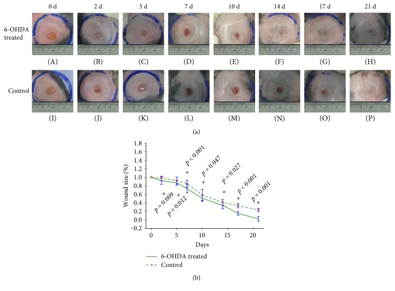
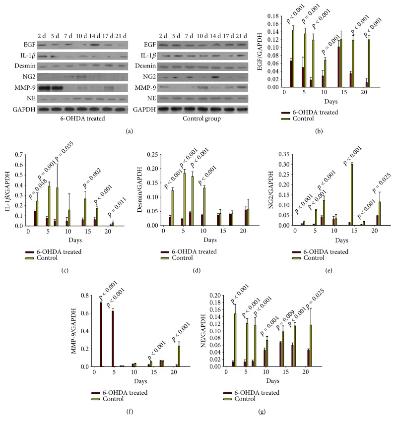
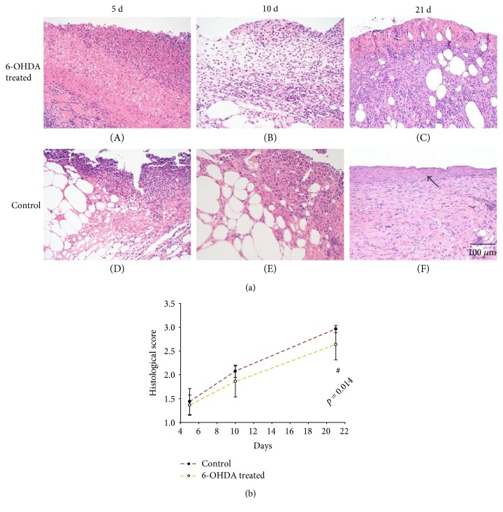
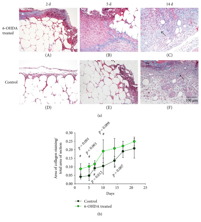
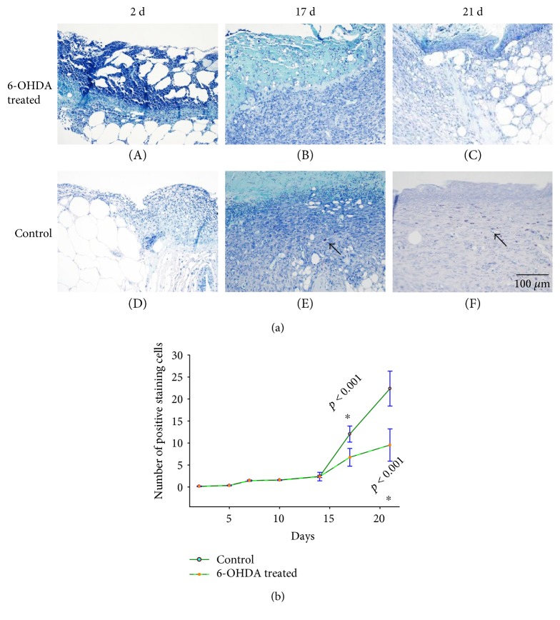
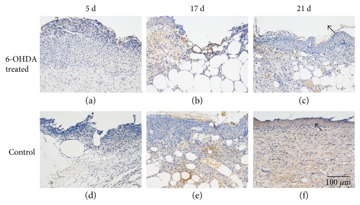
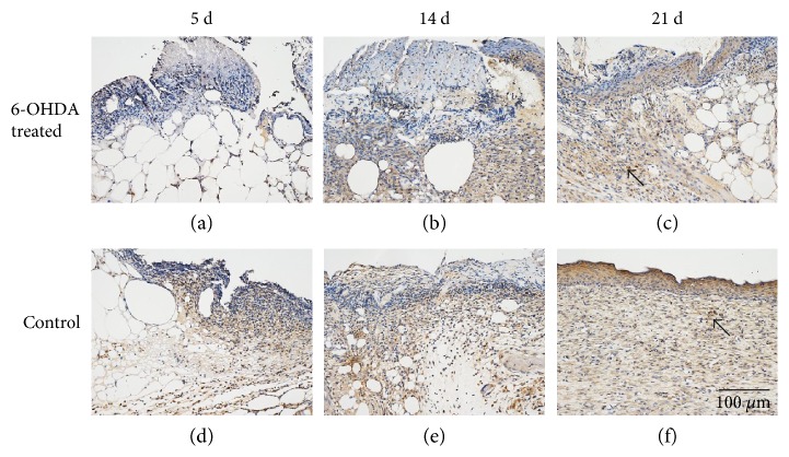
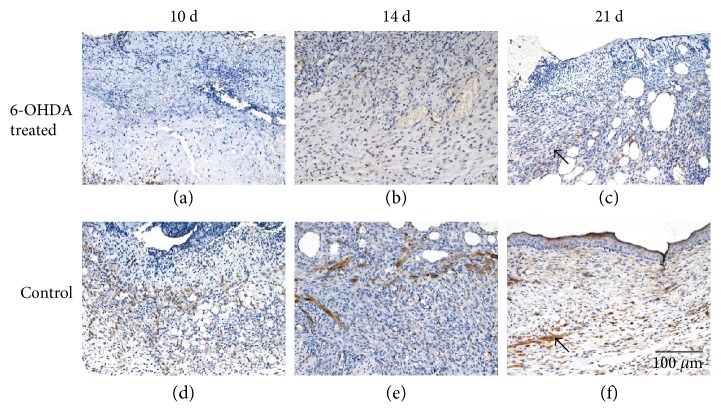
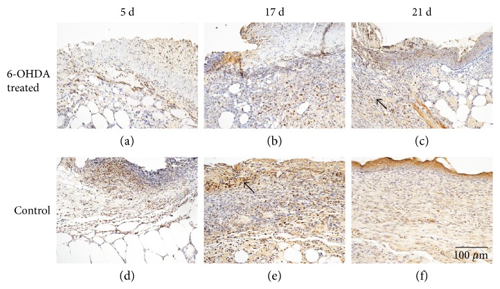
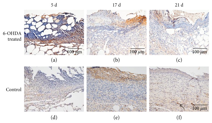
Previous studies focused on the effects of sympathetic denervation with 6-hydroxydopamine (6-OHDA) on nondiabetic wounds, but the effects of 6-OHDA on diabetic wounds have not been previously reported. In this study, treated mice received intraperitoneal 6-OHDA, and control mice received intraperitoneal injections of normal saline. Full-thickness wounds were established on the backs of mice. The wounds were sectioned (four mice per group) for analysis at 2, 5, 7, 10, 14, 17, and 21 days after injury. The wound areas in the control group were larger than those in the treatment group. Histological scores for epidermal and dermal regeneration were reduced in the 6-OHDA-treated group on day 21. The mast cells (MCs) in each field decreased after sympathectomy on days 17 and 21. The expression levels of norepinephrine, epidermal growth factor (EGF), interleukin-1 beta, NG2 proteoglycan, and desmin in the treatment group were less than those in the control group. In conclusion, 6-OHDA delays reepithelialization during wound healing in diabetic mice by decreasing EGF, but increases wound contraction by reducing IL-1 levels and the number of MCs. Besides, 6-OHDA led to reduced pericyte proliferation in diabetic wounds, which might explain the vascular dysfunction after sympathetic nerve loss in diabetic wounds.

摘要

先前的研究集中于去甲肾上腺素(6-OHDA)对非糖尿病伤口的交感神经切断作用,但 6-OHDA 对糖尿病伤口的影响尚未报道。在这项研究中,实验组小鼠接受腹腔内注射 6-OHDA,对照组小鼠接受腹腔内注射生理盐水。在小鼠背部建立全层伤口。在损伤后 2、5、7、10、14、17 和 21 天,每组 4 只小鼠对伤口进行切片分析。对照组的伤口面积大于治疗组。在第 21 天,治疗组表皮和真皮再生的组织学评分降低。在第 17 和 21 天交感神经切除后,每个视野中的肥大细胞(MC)减少。治疗组的去甲肾上腺素、表皮生长因子(EGF)、白细胞介素-1β、NG2 蛋白聚糖和结蛋白的表达水平低于对照组。总之,6-OHDA 通过降低 EGF 来延迟糖尿病小鼠伤口愈合中的再上皮化,但通过降低 IL-1 水平和 MC 数量来增加伤口收缩。此外,6-OHDA 导致糖尿病伤口中周细胞增殖减少,这可能解释了糖尿病伤口中交感神经丧失后的血管功能障碍。

https://cdn.ncbi.nlm.nih.gov/pmc/blobs/31e5/5632918/bd362525cb0a/JDR2017-7614685.010.jpg
https://cdn.ncbi.nlm.nih.gov/pmc/blobs/31e5/5632918/9bbe513c0b4d/JDR2017-7614685.001.jpg
https://cdn.ncbi.nlm.nih.gov/pmc/blobs/31e5/5632918/53f2a1c9102e/JDR2017-7614685.002.jpg
https://cdn.ncbi.nlm.nih.gov/pmc/blobs/31e5/5632918/26b143ad4e8e/JDR2017-7614685.003.jpg
https://cdn.ncbi.nlm.nih.gov/pmc/blobs/31e5/5632918/26a16b3e0dc5/JDR2017-7614685.004.jpg
https://cdn.ncbi.nlm.nih.gov/pmc/blobs/31e5/5632918/c1e9f074e914/JDR2017-7614685.005.jpg
https://cdn.ncbi.nlm.nih.gov/pmc/blobs/31e5/5632918/5d897d5be321/JDR2017-7614685.006.jpg
https://cdn.ncbi.nlm.nih.gov/pmc/blobs/31e5/5632918/9b9fcb95ee8d/JDR2017-7614685.007.jpg
https://cdn.ncbi.nlm.nih.gov/pmc/blobs/31e5/5632918/0f2fbac19cad/JDR2017-7614685.008.jpg
https://cdn.ncbi.nlm.nih.gov/pmc/blobs/31e5/5632918/8ceb6edeedcc/JDR2017-7614685.009.jpg
https://cdn.ncbi.nlm.nih.gov/pmc/blobs/31e5/5632918/bd362525cb0a/JDR2017-7614685.010.jpg

相似文献

1
Sympathetic Denervation Accelerates Wound Contraction but Inhibits Reepithelialization and Pericyte Proliferation in Diabetic Mice.交感神经切断加速糖尿病小鼠创面收缩,但抑制再上皮化和周细胞增殖。
J Diabetes Res. 2017;2017:7614685. doi: 10.1155/2017/7614685. Epub 2017 Sep 24.
2
Sympathetic denervation accelerates wound contraction but delays reepithelialization in rats.交感神经去神经支配会加速大鼠伤口收缩,但会延迟其上皮再形成。
Wound Repair Regen. 2005 Sep-Oct;13(5):498-505. doi: 10.1111/j.1067-1927.2005.00070.x.
3
Attenuation of Flightless I Increases Human Pericyte Proliferation, Migration and Angiogenic Functions and Improves Healing in Murine Diabetic Wounds.失活 Flightless I 可增强人周细胞的增殖、迁移和血管生成功能,并改善糖尿病小鼠创面的愈合。
Int J Mol Sci. 2020 Aug 5;21(16):5599. doi: 10.3390/ijms21165599.
4
Topical vascular endothelial growth factor accelerates diabetic wound healing through increased angiogenesis and by mobilizing and recruiting bone marrow-derived cells.局部应用血管内皮生长因子可通过增加血管生成以及动员和募集骨髓来源的细胞来加速糖尿病伤口愈合。
Am J Pathol. 2004 Jun;164(6):1935-47. doi: 10.1016/S0002-9440(10)63754-6.
5
Topical 1% propranolol cream promotes cutaneous wound healing in spontaneously diabetic mice.局部应用1%普萘洛尔乳膏可促进自发性糖尿病小鼠的皮肤伤口愈合。
Wound Repair Regen. 2017 May;25(3):389-397. doi: 10.1111/wrr.12546. Epub 2017 May 26.
6
Effects of chemical sympathectomy by means of 6-hydroxydopamine on insulin secretion and islet morphology in alloxan-diabetic mice.6-羟基多巴胺化学性交感神经切除术对四氧嘧啶糖尿病小鼠胰岛素分泌及胰岛形态的影响
Cell Tissue Res. 2002 Feb;307(2):203-9. doi: 10.1007/s00441-001-0496-5. Epub 2002 Jan 16.
7
Nerve growth factor accelerates wound healing in diabetic mice.神经生长因子可加速糖尿病小鼠的伤口愈合。
Wound Repair Regen. 2004 Jan-Feb;12(1):44-52. doi: 10.1111/j.1067-1927.2004.012110.x.
8
Decreased macrophage number and activation lead to reduced lymphatic vessel formation and contribute to impaired diabetic wound healing.巨噬细胞数量减少和活性降低导致淋巴管生成减少,并促使糖尿病伤口愈合受损。
Am J Pathol. 2007 Apr;170(4):1178-91. doi: 10.2353/ajpath.2007.060018.
9
Sympathetic denervation impairs epidermal healing in cutaneous wounds.
Wound Repair Regen. 1998 May-Jun;6(3):194-201. doi: 10.1046/j.1524-475x.1998.60305.x.
10
Targeting Imbalance between IL-1β and IL-1 Receptor Antagonist Ameliorates Delayed Epithelium Wound Healing in Diabetic Mouse Corneas.靶向白细胞介素-1β与白细胞介素-1受体拮抗剂之间的失衡可改善糖尿病小鼠角膜上皮伤口愈合延迟的情况。
Am J Pathol. 2016 Jun;186(6):1466-80. doi: 10.1016/j.ajpath.2016.01.019. Epub 2016 Apr 22.

引用本文的文献

1
Pericytes and Diabetic Microangiopathies: Tissue Resident Mesenchymal Stem Cells with High Plasticity and Regenerative Capacity.周细胞与糖尿病微血管病变:具有高可塑性和再生能力的组织驻留间充质干细胞
Int J Mol Sci. 2025 Jun 1;26(11):5333. doi: 10.3390/ijms26115333.
2
Advances in Zebrafish for Diabetes Mellitus with Wound Model.斑马鱼在糖尿病伤口模型研究中的进展
Bioengineering (Basel). 2023 Mar 6;10(3):330. doi: 10.3390/bioengineering10030330.
3
Sympathetic System in Wound Healing: Multistage Control in Normal and Diabetic Skin.

本文引用的文献

1
Angiogenesis and wound repair: when enough is enough.血管生成与伤口修复:适度即可。
J Leukoc Biol. 2016 Nov;100(5):979-984. doi: 10.1189/jlb.4MR0316-102R. Epub 2016 Jul 12.
2
Mast Cells Regulate Wound Healing in Diabetes.肥大细胞调节糖尿病中的伤口愈合。
Diabetes. 2016 Jul;65(7):2006-19. doi: 10.2337/db15-0340. Epub 2016 Apr 8.
3
Sympathetic nerves: How do they affect angiogenesis, particularly during wound healing of soft tissues?交感神经:它们如何影响血管生成,尤其是在软组织伤口愈合过程中?
创伤愈合中的交感神经系统:正常和糖尿病皮肤中的多阶段控制。
Int J Mol Sci. 2023 Jan 20;24(3):2045. doi: 10.3390/ijms24032045.
4
Sympathetic Denervation Alters the Inflammatory Response of Resident Muscularis Macrophages upon Surgical Trauma and Ameliorates Postoperative Ileus in Mice.交感神经切断术改变手术创伤后固有肌层巨噬细胞的炎症反应,并改善小鼠术后肠梗阻。
Int J Mol Sci. 2021 Jun 26;22(13):6872. doi: 10.3390/ijms22136872.
5
Cutaneous innervation in impaired diabetic wound healing.糖尿病伤口愈合受损中的皮肤神经支配
Transl Res. 2021 Oct;236:87-108. doi: 10.1016/j.trsl.2021.05.003. Epub 2021 May 23.
Clin Hemorheol Microcirc. 2016;62(2):181-91. doi: 10.3233/CH-152019.
4
Factors That Impair Wound Healing.影响伤口愈合的因素。
J Am Coll Clin Wound Spec. 2014 Mar 24;4(4):84-91. doi: 10.1016/j.jccw.2014.03.001. eCollection 2012 Dec.
5
The diabetic foot.糖尿病足
Diabetes Metab Syndr. 2016 Jan-Mar;10(1):48-60. doi: 10.1016/j.dsx.2015.04.002. Epub 2015 Apr 27.
6
Attempts to accelerate wound healing.加速伤口愈合的尝试。
J Dermatol Sci. 2014 Dec;76(3):169-72. doi: 10.1016/j.jdermsci.2014.11.001. Epub 2014 Nov 11.
7
The role of mast cells in cutaneous wound healing in streptozotocin-induced diabetic mice.肥大细胞在链脲佐菌素诱导的糖尿病小鼠皮肤伤口愈合中的作用。
Arch Dermatol Res. 2014 Nov;306(9):823-35. doi: 10.1007/s00403-014-1496-0. Epub 2014 Sep 14.
8
Epidermal Growth Factor and Epidermal Growth Factor Receptor: The Yin and Yang in the Treatment of Cutaneous Wounds and Cancer.表皮生长因子与表皮生长因子受体:皮肤创伤治疗与癌症治疗中的阴阳关系
Adv Wound Care (New Rochelle). 2013 Feb;2(1):24-29. doi: 10.1089/wound.2011.0326.
9
Exogenous bone morphogenetic protein-7 reduces hepatic fibrosis in Schistosoma japonicum-infected mice via transforming growth factor-β/Smad signaling.外源性骨形态发生蛋白 7 通过转化生长因子-β/Smad 信号通路减少日本血吸虫感染小鼠的肝纤维化。
World J Gastroenterol. 2013 Mar 7;19(9):1405-15. doi: 10.3748/wjg.v19.i9.1405.
10
Blocking interleukin-1β induces a healing-associated wound macrophage phenotype and improves healing in type 2 diabetes.阻断白细胞介素-1β可诱导与愈合相关的伤口巨噬细胞表型,并改善 2 型糖尿病的愈合。
Diabetes. 2013 Jul;62(7):2579-87. doi: 10.2337/db12-1450. Epub 2013 Mar 14.